7OUP | pdb_00007oup

Structure of human DPP3 in complex with a hydroxyethylene transition state peptidomimetic


Experimental Data Snapshot

  • Method: X-RAY DIFFRACTION
  • Resolution: 2.65 Å
  • R-Value Free: 
    0.253 (Depositor), 0.253 (DCC) 
  • R-Value Work: 
    0.197 (Depositor), 0.197 (DCC) 
  • R-Value Observed: 
    0.200 (Depositor) 

Starting Model: experimental
View more details

wwPDB Validation 3D Report Full Report

Validation slider image for 7OUP

This is version 2.2 of the entry. See complete history

Literature

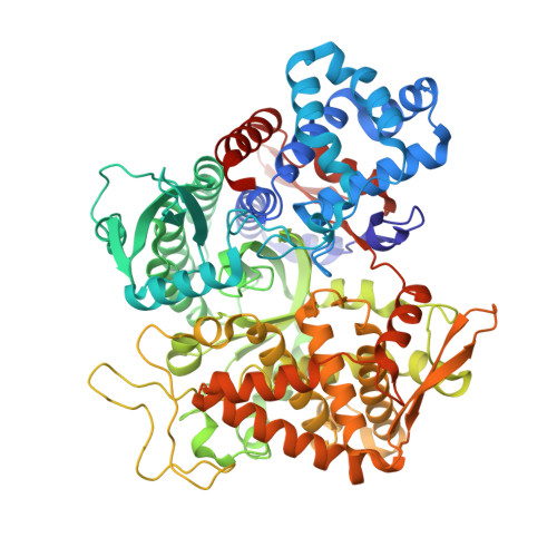
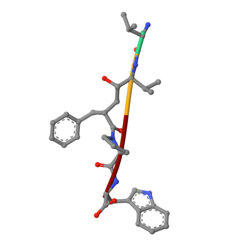
Efficient Entropy-Driven Inhibition of Dipeptidyl Peptidase III by Hydroxyethylene Transition-State Peptidomimetics.

Ivkovic, J.Jha, S.Lembacher-Fadum, C.Puschnig, J.Kumar, P.Reithofer, V.Gruber, K.Macheroux, P.Breinbauer, R.

(2021) Chemistry 27: 14108-14120

  • DOI: https://doi.org/10.1002/chem.202102204
  • Primary Citation Related Structures: 
    7OUP

  • PubMed Abstract: 

    Dipeptidyl peptidase III (DPP3) is a ubiquitously expressed Zn-dependent protease, which plays an important role in regulating endogenous peptide hormones, such as enkephalins or angiotensins. In previous biophysical studies, it could be shown that substrate binding is driven by a large entropic contribution due to the release of water molecules from the closing binding cleft. Here, the design, synthesis and biophysical characterization of peptidomimetic inhibitors is reported, using for the first time an hydroxyethylene transition-state mimetic for a metalloprotease. Efficient routes for the synthesis of both stereoisomers of the pseudopeptide core were developed, which allowed the synthesis of peptidomimetic inhibitors mimicking the VVYPW-motif of tynorphin. The best inhibitors inhibit DPP3 in the low μM range. Biophysical characterization by means of ITC measurement and X-ray crystallography confirm the unusual entropy-driven mode of binding. Stability assays demonstrated the desired stability of these inhibitors, which efficiently inhibited DPP3 in mouse brain homogenate.


  • Organizational Affiliation
    • Institute of Organic Chemistry, Graz University of Technology, Stremayrgasse 9, 8010, Graz, Austria.

Macromolecule Content 

  • Total Structure Weight: 83.39 kDa 
  • Atom Count: 5,870 
  • Modeled Residue Count: 728 
  • Deposited Residue Count: 741 
  • Unique protein chains: 2

Macromolecules

Find similar proteins by:|  3D Structure
Entity ID: 1
MoleculeChains  Sequence LengthOrganismDetailsImage
Dipeptidyl peptidase 3737Homo sapiensMutation(s): 1 
Gene Names: DPP3
EC: 3.4.14.4
UniProt & NIH Common Fund Data Resources
Find proteins for Q9NY33 (Homo sapiens)
Explore Q9NY33 
Go to UniProtKB:  Q9NY33
PHAROS:  Q9NY33
GTEx:  ENSG00000254986 
Entity Groups
Sequence Clusters30% Identity50% Identity70% Identity90% Identity95% Identity100% Identity
UniProt GroupQ9NY33
Sequence Annotations
Expand
Reference Sequence
Find similar proteins by:  Sequence   |   3D Structure  
Entity ID: 2
MoleculeChains  Sequence LengthOrganismDetailsImage
((2R,4S,5S)-5-((S)-2-amino-3-methylbutanamido)-2-benzyl-4-hydroxy-6-methylheptanoyl)-L-prolyl-L-tryptophanB [auth F]4synthetic constructMutation(s): 0 

Experimental Data & Validation

Experimental Data

  • Method: X-RAY DIFFRACTION
  • Resolution: 2.65 Å
  • R-Value Free:  0.253 (Depositor), 0.253 (DCC) 
  • R-Value Work:  0.197 (Depositor), 0.197 (DCC) 
  • R-Value Observed: 0.200 (Depositor) 
Space Group: C 1 2 1
Unit Cell:
Length ( Å )Angle ( ˚ )
a = 119.337α = 90
b = 105.597β = 93.651
c = 64.861γ = 90
Software Package:
Software NamePurpose
PHENIXrefinement
XDSdata reduction
XDSdata scaling
PHASERphasing

Structure Validation

View Full Validation Report


Entry History 

& Funding Information

Deposition Data


Funding OrganizationLocationGrant Number
Austrian Science FundAustriaW901

Revision History  (Full details and data files)

  • Version 1.0: 2021-08-11
    Type: Initial release
  • Version 1.1: 2021-10-20
    Changes: Data collection, Database references
  • Version 2.0: 2023-11-15
    Changes: Atomic model, Data collection, Derived calculations
  • Version 2.1: 2024-01-31
    Changes: Refinement description
  • Version 2.2: 2024-10-09
    Changes: Structure summary