Download Mendeley
Cryo-EM structures of pentameric autoinducer-2 exporter from Escherichia coli reveal its transport mechanism.
Khera, R., Mehdipour, A.R., Bolla, J.R., Kahnt, J., Welsch, S., Ermler, U., Muenke, C., Robinson, C.V., Hummer, G., Xie, H., Michel, H.(2022) EMBO J 41: e109990-e109990
- PubMed: 35698912 Search on PubMedSearch on PubMed Central
- DOI: https://doi.org/10.15252/embj.2021109990
- Primary Citation Related Structures:
7NB6, 7OT9 - PubMed Abstract:
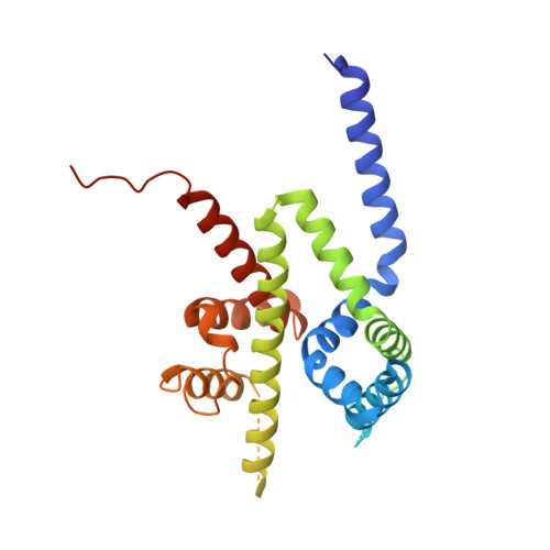
Bacteria utilize small extracellular molecules to communicate in order to collectively coordinate their behaviors in response to the population density. Autoinducer-2 (AI-2), a universal molecule for both intra- and inter-species communication, is involved in the regulation of biofilm formation, virulence, motility, chemotaxis, and antibiotic resistance. While many studies have been devoted to understanding the biosynthesis and sensing of AI-2, very little information is available on its export. The protein TqsA from Escherichia coli, which belongs to the AI-2 exporter superfamily, has been shown to export AI-2. Here, we report the cryogenic electron microscopic structures of two AI-2 exporters (TqsA and YdiK) from E. coli at 3.35 Å and 2.80 Å resolutions, respectively. Our structures suggest that the AI-2 exporter exists as a homo-pentameric complex. In silico molecular docking and native mass spectrometry experiments were employed to demonstrate the interaction between AI-2 and TqsA, and the results highlight the functional importance of two helical hairpins in substrate binding. We propose that each monomer works as an independent functional unit utilizing an elevator-type transport mechanism.
- Department of Molecular Membrane Biology, Max Planck Institute of Biophysics, Frankfurt am Main, Germany.
Organizational Affiliation: