7OS3 | pdb_00007os3

Crystal structure of Rhizobium etli inducible L-asparaginase ReAV solved by S-SAD (orthorhombic form START)


Experimental Data Snapshot

  • Method: X-RAY DIFFRACTION
  • Resolution: 2.18 Å
  • R-Value Free: 
    0.224 (Depositor), 0.234 (DCC) 
  • R-Value Work: 
    0.168 (Depositor), 0.177 (DCC) 

wwPDB Validation 3D Report Full Report

Validation slider image for 7OS3

This is version 1.2 of the entry. See complete history

Literature

Crystal structures of the elusive Rhizobium etli L-asparaginase reveal a peculiar active site.

Loch, J.I.Imiolczyk, B.Sliwiak, J.Wantuch, A.Bejger, M.Gilski, M.Jaskolski, M.

(2021) Nat Commun 12: 6717-6717

  • DOI: https://doi.org/10.1038/s41467-021-27105-x
  • Primary Citation Related Structures: 
    7OS3, 7OS5, 7OS6, 7OU1, 7OZ6

  • PubMed Abstract: 

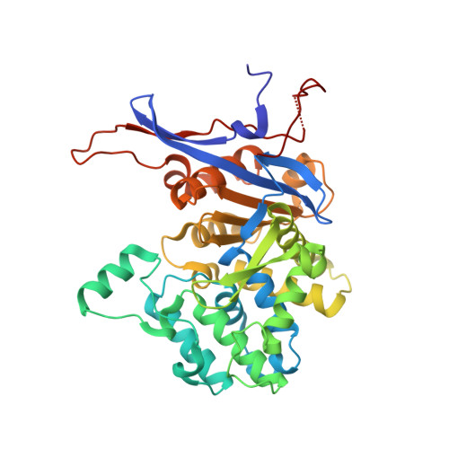
    Rhizobium etli, a nitrogen-fixing bacterial symbiont of legume plants, encodes an essential L-asparaginase (ReAV) with no sequence homology to known enzymes with this activity. High-resolution crystal structures of ReAV show indeed a structurally distinct, dimeric enzyme, with some resemblance to glutaminases and β-lactamases. However, ReAV has no glutaminase or lactamase activity, and at pH 9 its allosteric asparaginase activity is relatively high, with K m for L-Asn at 4.2 mM and k cat of 438 s -1 . The active site of ReAV, deduced from structural comparisons and confirmed by mutagenesis experiments, contains a highly specific Zn 2+ binding site without a catalytic role. The extensive active site includes residues with unusual chemical properties. There are two Ser-Lys tandems, all connected through a network of H-bonds to the Zn center, and three tightly bound water molecules near Ser48, which clearly indicate the catalytic nucleophile.


  • Organizational Affiliation
    • Department of Crystal Chemistry and Crystal Physics, Faculty of Chemistry, Jagiellonian University, Krakow, Poland.

Macromolecule Content 

  • Total Structure Weight: 79.31 kDa 
  • Atom Count: 5,639 
  • Modeled Residue Count: 718 
  • Deposited Residue Count: 746 
  • Unique protein chains: 1

Macromolecules

Find similar proteins by:|  3D Structure
Entity ID: 1
MoleculeChains  Sequence LengthOrganismDetailsImage
L-asparaginase II proteinA [auth AAA],
B [auth BBB]
373Rhizobium etli CFN 42Mutation(s): 0 
Gene Names: ansARHE_PE00350
UniProt
Find proteins for Q2K0Z2 (Rhizobium etli (strain ATCC 51251 / DSM 11541 / JCM 21823 / NBRC 15573 / CFN 42))
Explore Q2K0Z2 
Go to UniProtKB:  Q2K0Z2
Entity Groups
Sequence Clusters30% Identity50% Identity70% Identity90% Identity95% Identity100% Identity
UniProt GroupQ2K0Z2
Sequence Annotations
Expand
Reference Sequence

Experimental Data & Validation

Experimental Data

  • Method: X-RAY DIFFRACTION
  • Resolution: 2.18 Å
  • R-Value Free:  0.224 (Depositor), 0.234 (DCC) 
  • R-Value Work:  0.168 (Depositor), 0.177 (DCC) 
Space Group: P 21 21 21
Unit Cell:
Length ( Å )Angle ( ˚ )
a = 78.15α = 90
b = 91.05β = 90
c = 105.84γ = 90
Software Package:
Software NamePurpose
REFMACrefinement
XDSdata reduction
XSCALEdata scaling
HKL2Mapphasing

Structure Validation

View Full Validation Report


Entry History 

& Funding Information

Deposition Data


Funding OrganizationLocationGrant Number
Polish National Science CentrePoland2020/37/B/NZ1/03250

Revision History  (Full details and data files)

  • Version 1.0: 2021-11-24
    Type: Initial release
  • Version 1.1: 2021-12-01
    Changes: Database references
  • Version 1.2: 2024-06-19
    Changes: Data collection