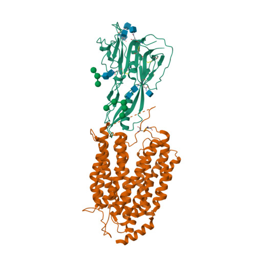
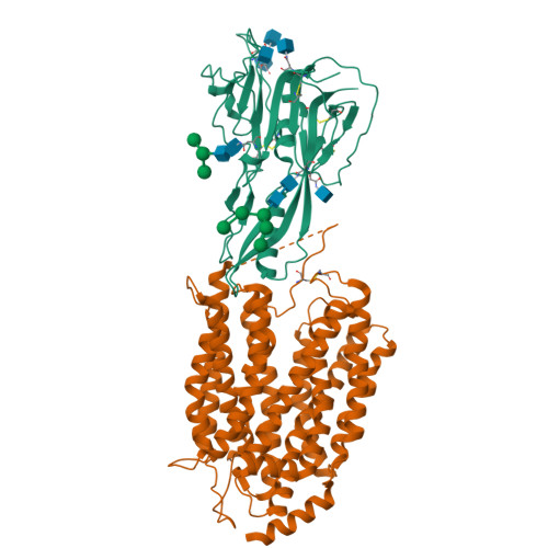
7OIX

Structure of thermostable human MFSD2A in complex with thermostable human Sync2


Experimental Data Snapshot

  • Method: ELECTRON MICROSCOPY
  • Resolution: 3.60 Å
  • Aggregation State: PARTICLE 
  • Reconstruction Method: SINGLE PARTICLE 

wwPDB Validation   3D Report Full Report


This is version 1.2 of the entry. See complete history


Literature

Structural insights into the lysophospholipid brain uptake mechanism and its inhibition by syncytin-2.

Martinez-Molledo, M.Nji, E.Reyes, N.

(2022) Nat Struct Mol Biol 29: 604-612

  • DOI: https://doi.org/10.1038/s41594-022-00786-8
  • Primary Citation of Related Structures:  
    7OIX

  • PubMed Abstract: 

    Brain development and function require uptake of essential omega-3 fatty acids in the form of lysophosphatidylcholine via major-facilitator superfamily transporter MFSD2A, a potential pharmaceutical target to modulate blood-brain barrier (BBB) permeability. MFSD2A is also the receptor of endogenous retroviral envelope syncytin-2 (SYNC2) in human placenta, where it mediates cell-cell fusion and formation of the maternal-fetal interface. Here, we report a cryo-electron microscopy structure of the human MFSD2A-SYNC2 complex that reveals a large hydrophobic cavity in the transporter C-terminal domain to occlude long aliphatic chains. The transporter architecture suggests an alternating-access transport mechanism for lipid substrates in mammalian MFS transporters. SYNC2 establishes an extensive binding interface with MFSD2A, and a SYNC2-soluble fragment acts as a long-sought-after inhibitor of MFSD2A transport. Our work uncovers molecular mechanisms important to brain and placenta development and function, and SYNC2-mediated inhibition of MFSD2A transport suggests strategies to aid delivery of therapeutic macromolecules across the BBB.


  • Organizational Affiliation

    Membrane Protein Mechanisms Group, European Institute of Chemistry and Biology, University of Bordeaux, CNRS-UMR5234, Bordeaux, France.


Macromolecules
Find similar proteins by:  (by identity cutoff)  |  3D Structure
Entity ID: 1
MoleculeChains Sequence LengthOrganismDetailsImage
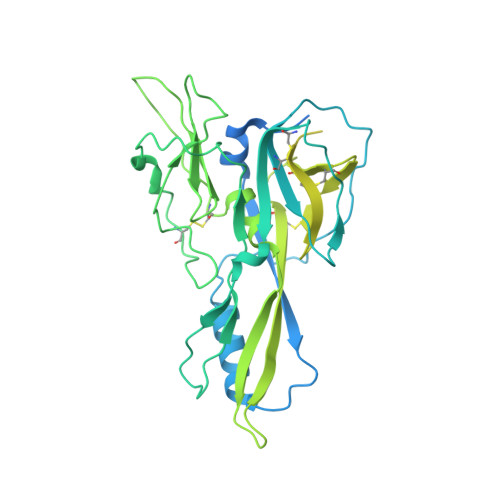
Syncytin-2538Homo sapiensMutation(s): 0 
Gene Names: ERVFRD-1ERVFRDE1UNQ6191/PRO20218
Membrane Entity: Yes 
UniProt & NIH Common Fund Data Resources
Find proteins for P60508 (Homo sapiens)
Explore P60508 
Go to UniProtKB:  P60508
PHAROS:  P60508
GTEx:  ENSG00000244476 
Entity Groups  
Sequence Clusters30% Identity50% Identity70% Identity90% Identity95% Identity100% Identity
UniProt GroupP60508
Glycosylation
Glycosylation Sites: 7Go to GlyGen: P60508-1
Sequence Annotations
Expand
  • Reference Sequence
Find similar proteins by:  (by identity cutoff)  |  3D Structure
Entity ID: 2
MoleculeChains Sequence LengthOrganismDetailsImage
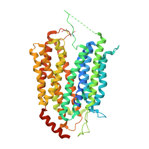
Isoform 2 of Sodium-dependent lysophosphatidylcholine symporter 1530Homo sapiensMutation(s): 0 
Gene Names: MFSD2AMFSD2NLS1HMFN0656PP9177UNQ300/PRO341
Membrane Entity: Yes 
UniProt & NIH Common Fund Data Resources
Find proteins for Q8NA29 (Homo sapiens)
Explore Q8NA29 
Go to UniProtKB:  Q8NA29
PHAROS:  Q8NA29
GTEx:  ENSG00000168389 
Entity Groups  
Sequence Clusters30% Identity50% Identity70% Identity90% Identity95% Identity100% Identity
UniProt GroupQ8NA29
Sequence Annotations
Expand
  • Reference Sequence
Oligosaccharides

Help

Entity ID: 3
MoleculeChains Length2D Diagram Glycosylation3D Interactions
alpha-D-mannopyranose-(1-3)-[alpha-D-mannopyranose-(1-6)]beta-D-mannopyranose-(1-6)-2-acetamido-2-deoxy-beta-D-glucopyranose-(1-4)-2-acetamido-2-deoxy-beta-D-glucopyranose
C
5N-Glycosylation
Entity ID: 4
MoleculeChains Length2D Diagram Glycosylation3D Interactions
alpha-D-mannopyranose-(1-2)-alpha-D-mannopyranose-(1-3)-[alpha-D-mannopyranose-(1-6)-alpha-D-mannopyranose-(1-6)]beta-D-mannopyranose-(1-4)-2-acetamido-2-deoxy-beta-D-glucopyranose-(1-4)-2-acetamido-2-deoxy-beta-D-glucopyranose
D
7N-Glycosylation
Glycosylation Resources
GlyTouCan:  G23799GS
GlyCosmos:  G23799GS
GlyGen:  G23799GS
Experimental Data & Validation

Experimental Data

  • Method: ELECTRON MICROSCOPY
  • Resolution: 3.60 Å
  • Aggregation State: PARTICLE 
  • Reconstruction Method: SINGLE PARTICLE 

Structure Validation

View Full Validation Report


Entry History & Funding Information

Deposition Data


Funding OrganizationLocationGrant Number
European Research Council (ERC)European Union771965

Revision History  (Full details and data files)

  • Version 1.0: 2022-06-01
    Type: Initial release
  • Version 1.1: 2022-06-29
    Changes: Database references
  • Version 1.2: 2024-10-16
    Changes: Data collection, Structure summary