7OIH | pdb_00007oih

Glycosylation in the crystal structure of neutrophil myeloperoxidase


Experimental Data Snapshot

  • Method: X-RAY DIFFRACTION
  • Resolution: 2.60 Å
  • R-Value Free: 
    0.220 (Depositor), 0.191 (DCC) 
  • R-Value Work: 
    0.179 (Depositor) 

Starting Model: experimental
View more details

wwPDB Validation 3D Report Full Report

Validation slider image for 7OIH

Ligand Structure Quality Assessment 


This is version 1.3 of the entry. See complete history

Literature

Native glycosylation and binding of the antidepressant paroxetine in a low-resolution crystal structure of human myeloperoxidase.

Krawczyk, L.Semwal, S.Soubhye, J.Lemri Ouadriri, S.Prevost, M.Van Antwerpen, P.Roos, G.Bouckaert, J.

(2022) Acta Crystallogr D Struct Biol 78: 1099-1109

  • DOI: https://doi.org/10.1107/S2059798322007082
  • Primary Citation Related Structures: 
    7OIH

  • PubMed Abstract: 

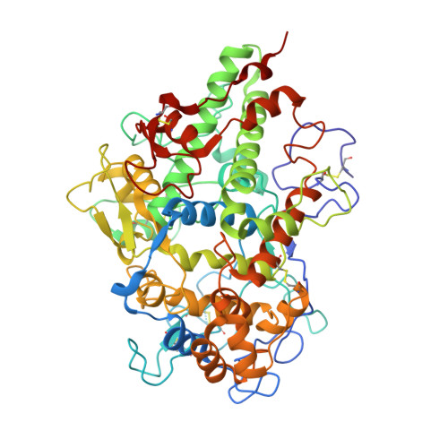
    Human myeloperoxidase (MPO) utilizes hydrogen peroxide to oxidize organic compounds and as such plays an essential role in cell-component synthesis, in metabolic and elimination pathways, and in the front-line defence against pathogens. Moreover, MPO is increasingly being reported to play a role in inflammation. The enzymatic activity of MPO has also been shown to depend on its glycosylation. Mammalian MPO crystal structures deposited in the Protein Data Bank (PDB) present only a partial identification of their glycosylation. Here, a newly obtained crystal structure of MPO containing four disulfide-linked dimers and showing an elaborate collection of glycans is reported. These are compared with the glycans identified in proteomics studies and from 18 human MPO structures available in the PDB. The crystal structure also contains bound paroxetine, a blocker of serotonin reuptake that has previously been identified as an irreversible inhibitor of MPO, in the presence of thiocyanate, a physiological substrate of MPO.


  • Organizational Affiliation
    • UGSF, CNRS, 50 Avenue de Halley, 59658 Villeneuve d'Ascq, France.

Macromolecule Content 

  • Total Structure Weight: 558.96 kDa 
  • Atom Count: 41,050 
  • Modeled Residue Count: 4,567 
  • Deposited Residue Count: 4,632 
  • Unique protein chains: 1

Macromolecules

Find similar proteins by:|  3D Structure
Entity ID: 1
MoleculeChains  Sequence LengthOrganismDetailsImage
Myeloperoxidase
A, B, C, D, E
A, B, C, D, E, F, G, H
579Homo sapiensMutation(s): 0 
EC: 1.11.2.2
UniProt & NIH Common Fund Data Resources
Find proteins for P05164 (Homo sapiens)
Explore P05164 
Go to UniProtKB:  P05164
PHAROS:  P05164
GTEx:  ENSG00000005381 
Entity Groups
Sequence Clusters30% Identity50% Identity70% Identity90% Identity95% Identity100% Identity
UniProt GroupP05164
Glycosylation
Glycosylation Sites: 4Go to GlyGen: P05164-1
Sequence Annotations
Expand
Reference Sequence

Oligosaccharides

Help  
Entity ID: 2
MoleculeChains Length2D Diagram GlycosylationD Interactions
beta-D-mannopyranose-(1-4)-2-acetamido-2-deoxy-beta-D-glucopyranose-(1-4)-2-acetamido-2-deoxy-beta-D-glucopyranoseCA [auth b],
EA [auth e],
I,
J,
Z [auth Y]
3N-Glycosylation
Glycosylation Resources
GlyTouCan: G15407YE
GlyCosmos: G15407YE
GlyGen: G15407YE
Entity ID: 3
MoleculeChains Length2D Diagram GlycosylationD Interactions
2-acetamido-2-deoxy-beta-D-glucopyranose-(1-2)-alpha-D-mannopyranose-(1-6)-[alpha-D-mannopyranose-(1-3)]beta-D-mannopyranose-(1-4)-2-acetamido-2-deoxy-beta-D-glucopyranose-(1-4)-[alpha-L-fucopyranose-(1-6)]2-acetamido-2-deoxy-beta-D-glucopyranoseK [auth c],
L [auth g],
N [auth L]
7N-Glycosylation
Glycosylation Resources
GlyTouCan: G45889JQ
GlyCosmos: G45889JQ
GlyGen: G45889JQ
Entity ID: 4
MoleculeChains Length2D Diagram GlycosylationD Interactions
alpha-D-mannopyranose-(1-3)-alpha-D-mannopyranose-(1-6)-[alpha-D-mannopyranose-(1-3)]beta-D-mannopyranose-(1-4)-2-acetamido-2-deoxy-beta-D-glucopyranose-(1-4)-2-acetamido-2-deoxy-beta-D-glucopyranoseM [auth K],
T [auth R]
6N-Glycosylation
Glycosylation Resources
GlyTouCan: G09724ZC
GlyCosmos: G09724ZC
GlyGen: G09724ZC
Entity ID: 5
MoleculeChains Length2D Diagram GlycosylationD Interactions
2-acetamido-2-deoxy-beta-D-glucopyranose-(1-4)-2-acetamido-2-deoxy-beta-D-glucopyranose
DA [auth d],
FA [auth f],
GA [auth S],
O [auth M],
Q [auth O],
DA [auth d],
FA [auth f],
GA [auth S],
O [auth M],
Q [auth O],
W [auth V]
2N-Glycosylation
Glycosylation Resources
GlyTouCan: G42666HT
GlyCosmos: G42666HT
GlyGen: G42666HT
Entity ID: 6
MoleculeChains Length2D Diagram GlycosylationD Interactions
alpha-D-mannopyranose-(1-3)-[alpha-D-mannopyranose-(1-6)]beta-D-mannopyranose-(1-4)-2-acetamido-2-deoxy-beta-D-glucopyranose-(1-4)-[alpha-L-fucopyranose-(1-6)]2-acetamido-2-deoxy-beta-D-glucopyranoseAA [auth Z],
P [auth N],
S [auth Q],
U [auth T],
X [auth W]
6N-Glycosylation
Glycosylation Resources
GlyTouCan: G82348BZ
GlyCosmos: G82348BZ
GlyGen: G82348BZ
Entity ID: 7
MoleculeChains Length2D Diagram GlycosylationD Interactions
alpha-D-mannopyranose-(1-3)-beta-D-mannopyranose-(1-4)-2-acetamido-2-deoxy-beta-D-glucopyranose-(1-4)-2-acetamido-2-deoxy-beta-D-glucopyranoseR [auth P]4N/A
Glycosylation Resources
GlyTouCan: G81315DD
GlyCosmos: G81315DD
GlyGen: G81315DD
Entity ID: 8
MoleculeChains Length2D Diagram GlycosylationD Interactions
alpha-D-mannopyranose-(1-6)-beta-D-mannopyranose-(1-4)-2-acetamido-2-deoxy-beta-D-glucopyranose-(1-4)-2-acetamido-2-deoxy-beta-D-glucopyranoseV [auth U],
Y [auth X]
4N-Glycosylation
Glycosylation Resources
GlyTouCan: G22573RC
GlyCosmos: G22573RC
GlyGen: G22573RC
Entity ID: 9
MoleculeChains Length2D Diagram GlycosylationD Interactions
alpha-D-mannopyranose-(1-2)-alpha-D-mannopyranose-(1-3)-[alpha-D-mannopyranose-(1-6)]beta-D-mannopyranose-(1-4)-2-acetamido-2-deoxy-beta-D-glucopyranose-(1-4)-2-acetamido-2-deoxy-beta-D-glucopyranoseBA [auth a]6N-Glycosylation
Glycosylation Resources
GlyTouCan: G56014GC
GlyCosmos: G56014GC
GlyGen: G56014GC

Small Molecules

Ligands 7 Unique
IDChains Name / Formula / InChI Key2D Diagram3D Interactions
HEM
(Subject of Investigation/LOI)

Query on HEM


Download:Ideal Coordinates CCD File
GC [auth E]
ID [auth H]
JB [auth C]
LA [auth A]
OC [auth F]
GC [auth E],
ID [auth H],
JB [auth C],
LA [auth A],
OC [auth F],
TB [auth D],
XA [auth B],
ZC [auth G]
PROTOPORPHYRIN IX CONTAINING FE
C34 H32 Fe N4 O4
KABFMIBPWCXCRK-RGGAHWMASA-L
8PR
(Subject of Investigation/LOI)

Query on 8PR


Download:Ideal Coordinates CCD File
KD [auth H],
NA [auth A],
QC [auth F],
VB [auth D]
Paroxetine
C19 H20 F N O3
AHOUBRCZNHFOSL-YOEHRIQHSA-N
NAG
(Subject of Investigation/LOI)

Query on NAG


Download:Ideal Coordinates CCD File
HB [auth C],
IB [auth C],
SB [auth D],
VA [auth B],
WA [auth B]
2-acetamido-2-deoxy-beta-D-glucopyranose
C8 H15 N O6
OVRNDRQMDRJTHS-FMDGEEDCSA-N
PO4

Query on PO4


Download:Ideal Coordinates CCD File
AC [auth D]
BC [auth D]
CB [auth B]
CD [auth G]
DB [auth B]
AC [auth D],
BC [auth D],
CB [auth B],
CD [auth G],
DB [auth B],
DD [auth G],
JC [auth E],
MD [auth H],
NB [auth C],
ND [auth H],
OB [auth C],
QA [auth A],
TC [auth F],
ZB [auth D]
PHOSPHATE ION
O4 P
NBIIXXVUZAFLBC-UHFFFAOYSA-K
SCN

Query on SCN


Download:Ideal Coordinates CCD File
AD [auth G]
HC [auth E]
JD [auth H]
KB [auth C]
MA [auth A]
AD [auth G],
HC [auth E],
JD [auth H],
KB [auth C],
MA [auth A],
PB [auth D],
PC [auth F],
UB [auth D],
YA [auth B],
ZA [auth B]
THIOCYANATE ION
C N S
ZMZDMBWJUHKJPS-UHFFFAOYSA-M
CA

Query on CA


Download:Ideal Coordinates CCD File
FC [auth E]
GB [auth C]
HD [auth H]
KA [auth A]
MC [auth F]
FC [auth E],
GB [auth C],
HD [auth H],
KA [auth A],
MC [auth F],
RB [auth D],
UA [auth B],
YC [auth G]
CALCIUM ION
Ca
BHPQYMZQTOCNFJ-UHFFFAOYSA-N
CL

Query on CL


Download:Ideal Coordinates CCD File
AB [auth B]
BB [auth B]
BD [auth G]
CC [auth E]
DC [auth E]
AB [auth B],
BB [auth B],
BD [auth G],
CC [auth E],
DC [auth E],
EB [auth C],
EC [auth E],
ED [auth H],
FB [auth C],
FD [auth H],
GD [auth H],
HA [auth A],
IA [auth A],
IC [auth E],
JA [auth A],
KC [auth F],
LB [auth C],
LC [auth F],
LD [auth H],
MB [auth C],
NC [auth F],
OA [auth A],
PA [auth A],
QB [auth D],
RA [auth B],
RC [auth F],
SA [auth B],
SC [auth F],
TA [auth B],
UC [auth G],
VC [auth G],
WB [auth D],
WC [auth G],
XB [auth D],
XC [auth G],
YB [auth D]
CHLORIDE ION
Cl
VEXZGXHMUGYJMC-UHFFFAOYSA-M
Modified Residues  1 Unique
IDChains TypeFormula2D DiagramParent
CSO
Query on CSO
A, B, C, D, E
A, B, C, D, E, F, G, H
L-PEPTIDE LINKINGC3 H7 N O3 SCYS
Binding Affinity Annotations 
IDSourceBinding Affinity
8PR BindingDB:  7OIH IC50: 20 (nM) from 1 assay(s)

Experimental Data & Validation

Experimental Data

  • Method: X-RAY DIFFRACTION
  • Resolution: 2.60 Å
  • R-Value Free:  0.220 (Depositor), 0.191 (DCC) 
  • R-Value Work:  0.179 (Depositor) 
Space Group: C 1 2 1
Unit Cell:
Length ( Å )Angle ( ˚ )
a = 155.91α = 90
b = 144.634β = 91.526
c = 236.454γ = 90
Software Package:
Software NamePurpose
REFMACrefinement
PHENIXrefinement
XDSdata reduction
SCALAdata scaling
PHASERphasing

Structure Validation

View Full Validation Report


Ligand Structure Quality Assessment 


Entry History 

& Funding Information

Deposition Data


Funding OrganizationLocationGrant Number
H2020 Marie Curie Actions of the European CommissionEuropean Union847568

Revision History  (Full details and data files)

  • Version 1.0: 2022-08-24
    Type: Initial release
  • Version 1.1: 2022-09-14
    Changes: Database references
  • Version 1.2: 2024-01-31
    Changes: Data collection, Refinement description
  • Version 1.3: 2024-11-06
    Changes: Structure summary