Equilibria between conformational states of the Ras oncogene protein revealed by high pressure crystallography.
Girard, E., Lopes, P., Spoerner, M., Dhaussy, A.C., Prange, T., Kalbitzer, H.R., Colloc'h, N.(2022) Chem Sci 13: 2001-2010
- PubMed: 35308861
- DOI: https://doi.org/10.1039/d1sc05488k
- Primary Citation of Related Structures:
7OG9, 7OGA, 7OGB, 7OGC, 7OGD, 7OGE, 7OGF - PubMed Abstract:
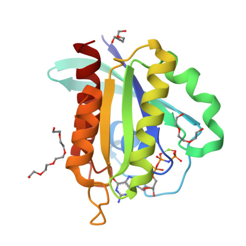
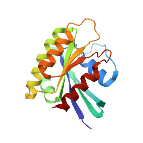
In this work, we experimentally investigate the allosteric transitions between conformational states on the Ras oncogene protein using high pressure crystallography. Ras protein is a small GTPase involved in central regulatory processes occurring in multiple conformational states. Ras acts as a molecular switch between active GTP-bound, and inactive GDP-bound states, controlling essential signal transduction pathways. An allosteric network of interactions between the effector binding regions and the membrane interacting regions is involved in Ras cycling. The conformational states which coexist simultaneously in solution possess higher Gibbs free energy than the ground state. Equilibria between these states can be shifted by applying pressure favouring conformations with lower partial molar volume, and has been previously analyzed by high-pressure NMR spectroscopy. High-pressure macromolecular crystallography (HPMX) is a powerful tool perfectly complementary to high-pressure NMR, allowing characterization at the molecular level with a high resolution the different allosteric states involved in the Ras cycling. We observe a transition above 300 MPa in the crystal leading to more stable conformers. Thus, we compare the crystallographic structures of Ras(wt)·Mg 2+ ·GppNHp and Ras(D33K)·Mg 2+ ·GppNHp at various high hydrostatic pressures. This gives insight into per-residue descriptions of the structural plasticity involved in allosteric equilibria between conformers. We have mapped out at atomic resolution the different segments of Ras protein which remain in the ground-state conformation or undergo structural changes, adopting excited-energy conformations corresponding to transient intermediate states. Such in crystallo phase transitions induced by pressure open the possibility to finely explore the structural determinants related to switching between Ras allosteric sub-states without any mutations nor exogenous partners.
Organizational Affiliation:
Univ. Grenoble Alpes, CEA, CNRS, IBS Grenoble France.


