7ODK | pdb_00007odk

Plant peptide hormone receptor H1


Experimental Data Snapshot

  • Method: X-RAY DIFFRACTION
  • Resolution: 1.83 Å
  • R-Value Free: 
    0.216 (Depositor), 0.226 (DCC) 
  • R-Value Work: 
    0.204 (Depositor), 0.212 (DCC) 

Starting Model: experimental
View more details

wwPDB Validation 3D Report Full Report

Validation slider image for 7ODK

Ligand Structure Quality Assessment 


This is version 1.3 of the entry. See complete history

Literature

HSL1 and BAM1/2 impact epidermal cell development by sensing distinct signaling peptides.

Roman, A.O.Jimenez-Sandoval, P.Augustin, S.Broyart, C.Hothorn, L.A.Santiago, J.

(2022) Nat Commun 13: 876-876

  • DOI: https://doi.org/10.1038/s41467-022-28558-4
  • Primary Citation Related Structures: 
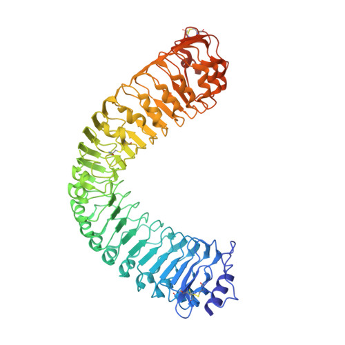
    7ODK, 7ODV, 7OGO, 7OGQ, 7OGU, 7OGZ

  • PubMed Abstract: 

    The membrane receptor kinases HAESA and HSL2 recognize a family of IDA/IDL signaling peptides to control cell separation processes in different plant organs. The homologous HSL1 has been reported to regulate epidermal cell patterning by interacting with a different class of signaling peptides from the CLE family. Here we demonstrate that HSL1 binds IDA/IDL peptides with high, and CLE peptides with lower affinity, respectively. Ligand sensing capability and receptor activation of HSL1 require a SERK co-receptor kinase. Crystal structures with IDA/IDLs or with CLE9 reveal that HSL1-SERK1 complex recognizes the entire IDA/IDL signaling peptide, while only parts of CLE9 are bound to the receptor. In contrast, the receptor kinase BAM1 interacts with the entire CLE9 peptide with high affinity and specificity. Furthermore, the receptor tandem BAM1/BAM2 regulates epidermal cell division homeostasis. Consequently, HSL1-IDLs and BAM1/BAM2-CLEs independently regulate cell patterning in the leaf epidermal tissue.


  • Organizational Affiliation
    • The Plant Signaling Mechanisms Laboratory, Department of Plant Molecular Biology, University of Lausanne, 1015, Lausanne, Switzerland.

Macromolecule Content 

  • Total Structure Weight: 142.98 kDa 
  • Atom Count: 10,222 
  • Modeled Residue Count: 1,174 
  • Deposited Residue Count: 1,234 
  • Unique protein chains: 1

Macromolecules

Find similar proteins by:|  3D Structure
Entity ID: 1
MoleculeChains  Sequence LengthOrganismDetailsImage
Receptor-like protein kinase HSL1A [auth AAA],
B [auth BBB]
617Arabidopsis thalianaMutation(s): 0 
Gene Names: HSL1At1g28440F3M18.12
EC: 2.7.11.1
UniProt
Find proteins for Q9SGP2 (Arabidopsis thaliana)
Explore Q9SGP2 
Go to UniProtKB:  Q9SGP2
Entity Groups
Sequence Clusters30% Identity50% Identity70% Identity90% Identity95% Identity100% Identity
UniProt GroupQ9SGP2
Glycosylation
Glycosylation Sites: 10
Sequence Annotations
Expand
Reference Sequence

Oligosaccharides

Help  
Entity ID: 2
MoleculeChains Length2D Diagram GlycosylationD Interactions
beta-D-mannopyranose-(1-4)-2-acetamido-2-deoxy-beta-D-glucopyranose-(1-4)-2-acetamido-2-deoxy-beta-D-glucopyranoseC [auth AlA],
J [auth BkB]
3N-Glycosylation
Glycosylation Resources
GlyTouCan: G15407YE
GlyCosmos: G15407YE
GlyGen: G15407YE
Entity ID: 3
MoleculeChains Length2D Diagram GlycosylationD Interactions
2-acetamido-2-deoxy-beta-D-glucopyranose-(1-4)-2-acetamido-2-deoxy-beta-D-glucopyranose
D [auth AmA],
E [auth AnA],
F [auth AoA],
G [auth AqA],
K [auth BmB],
D [auth AmA],
E [auth AnA],
F [auth AoA],
G [auth AqA],
K [auth BmB],
L [auth BoB],
M [auth BpB],
O [auth BvB],
P [auth BwB]
2N-Glycosylation
Glycosylation Resources
GlyTouCan: G42666HT
GlyCosmos: G42666HT
GlyGen: G42666HT
Entity ID: 4
MoleculeChains Length2D Diagram GlycosylationD Interactions
alpha-L-fucopyranose-(1-3)-[2-acetamido-2-deoxy-beta-D-glucopyranose-(1-4)][alpha-L-fucopyranose-(1-6)]2-acetamido-2-deoxy-beta-D-glucopyranoseH [auth AsA],
N [auth BqB]
4N-Glycosylation
Glycosylation Resources
GlyTouCan: G63564LA
GlyCosmos: G63564LA
GlyGen: G63564LA
Entity ID: 5
MoleculeChains Length2D Diagram GlycosylationD Interactions
alpha-L-fucopyranose-(1-3)-[2-acetamido-2-deoxy-beta-D-glucopyranose-(1-4)]2-acetamido-2-deoxy-beta-D-glucopyranoseI [auth AwA]3N-Glycosylation
Glycosylation Resources
GlyTouCan: G11971MR
GlyCosmos: G11971MR
GlyGen: G11971MR

Small Molecules

Ligands 4 Unique
IDChains Name / Formula / InChI Key2D Diagram3D Interactions
NAG

Query on NAG


Download:Ideal Coordinates CCD File
BA [auth AAA]
CA [auth AAA]
DA [auth AAA]
PA [auth BBB]
QA [auth BBB]
BA [auth AAA],
CA [auth AAA],
DA [auth AAA],
PA [auth BBB],
QA [auth BBB],
RA [auth BBB]
2-acetamido-2-deoxy-beta-D-glucopyranose
C8 H15 N O6
OVRNDRQMDRJTHS-FMDGEEDCSA-N
SO4

Query on SO4


Download:Ideal Coordinates CCD File
EA [auth AAA],
SA [auth BBB]
SULFATE ION
O4 S
QAOWNCQODCNURD-UHFFFAOYSA-L
EDO

Query on EDO


Download:Ideal Coordinates CCD File
AA [auth AAA]
FA [auth BBB]
GA [auth BBB]
HA [auth BBB]
IA [auth BBB]
AA [auth AAA],
FA [auth BBB],
GA [auth BBB],
HA [auth BBB],
IA [auth BBB],
JA [auth BBB],
KA [auth BBB],
LA [auth BBB],
MA [auth BBB],
NA [auth BBB],
OA [auth BBB],
Q [auth AAA],
R [auth AAA],
S [auth AAA],
T [auth AAA],
U [auth AAA],
V [auth AAA],
W [auth AAA],
X [auth AAA],
Y [auth AAA],
Z [auth AAA]
1,2-ETHANEDIOL
C2 H6 O2
LYCAIKOWRPUZTN-UHFFFAOYSA-N
NA

Query on NA


Download:Ideal Coordinates CCD File
TA [auth BBB]SODIUM ION
Na
FKNQFGJONOIPTF-UHFFFAOYSA-N

Experimental Data & Validation

Experimental Data

  • Method: X-RAY DIFFRACTION
  • Resolution: 1.83 Å
  • R-Value Free:  0.216 (Depositor), 0.226 (DCC) 
  • R-Value Work:  0.204 (Depositor), 0.212 (DCC) 
Space Group: P 1
Unit Cell:
Length ( Å )Angle ( ˚ )
a = 77.539α = 99.178
b = 84.001β = 113.727
c = 89.061γ = 108.41
Software Package:
Software NamePurpose
REFMACrefinement
REFMACrefinement
Aimlessdata scaling
XDSdata reduction
Cootmodel building
PHASERphasing

Structure Validation

View Full Validation Report


Ligand Structure Quality Assessment 


Entry History 

& Funding Information

Deposition Data


Funding OrganizationLocationGrant Number
Swiss National Science FoundationSwitzerland31003A_173101

Revision History  (Full details and data files)

  • Version 1.0: 2022-02-16
    Type: Initial release
  • Version 1.1: 2022-03-02
    Changes: Database references
  • Version 1.2: 2024-01-31
    Changes: Data collection, Derived calculations, Refinement description
  • Version 1.3: 2024-11-20
    Changes: Structure summary