7OD9 | pdb_00007od9

Crystal structure of activated CheY fused to the C-terminal domain of CheF


Experimental Data Snapshot

  • Method: X-RAY DIFFRACTION
  • Resolution: 2.30 Å
  • R-Value Free: 
    0.307 (Depositor), 0.308 (DCC) 
  • R-Value Work: 
    0.249 (Depositor), 0.250 (DCC) 
  • R-Value Observed: 
    0.252 (Depositor) 

Starting Model: experimental
View more details

wwPDB Validation 3D Report Full Report

Validation slider image for 7OD9

This is version 1.2 of the entry. See complete history

Literature

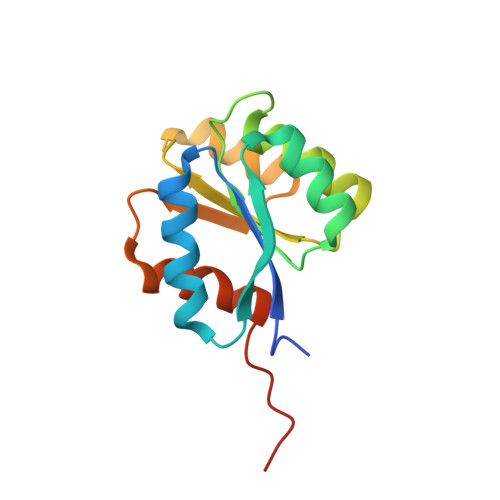
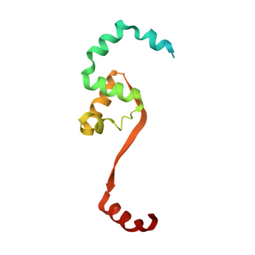
Structural insights into the mechanism of archaellar rotational switching.

Altegoer, F.Quax, T.E.F.Weiland, P.Nussbaum, P.Giammarinaro, P.I.Patro, M.Li, Z.Oesterhelt, D.Grininger, M.Albers, S.V.Bange, G.

(2022) Nat Commun 13: 2857-2857

  • DOI: https://doi.org/10.1038/s41467-022-30358-9
  • Primary Citation Related Structures: 
    7OD9, 7OVP

  • PubMed Abstract: 

    Signal transduction via phosphorylated CheY towards the flagellum and the archaellum involves a conserved mechanism of CheY phosphorylation and subsequent conformational changes within CheY. This mechanism is conserved among bacteria and archaea, despite substantial differences in the composition and architecture of archaellum and flagellum, respectively. Phosphorylated CheY has higher affinity towards the bacterial C-ring and its binding leads to conformational changes in the flagellar motor and subsequent rotational switching of the flagellum. In archaea, the adaptor protein CheF resides at the cytoplasmic face of the archaeal C-ring formed by the proteins ArlCDE and interacts with phosphorylated CheY. While the mechanism of CheY binding to the C-ring is well-studied in bacteria, the role of CheF in archaea remains enigmatic and mechanistic insights are absent. Here, we have determined the atomic structures of CheF alone and in complex with activated CheY by X-ray crystallography. CheF forms an elongated dimer with a twisted architecture. We show that CheY binds to the C-terminal tail domain of CheF leading to slight conformational changes within CheF. Our structural, biochemical and genetic analyses reveal the mechanistic basis for CheY binding to CheF and allow us to propose a model for rotational switching of the archaellum.


  • Organizational Affiliation
    • Philipps-University Marburg, Center for Synthetic Microbiology (SYNMIKRO) & Faculty of Chemistry, Karl-von-Frisch-Straße 14, 35043, Marburg, Germany. altegoer@hhu.de.

Macromolecule Content 

  • Total Structure Weight: 56.79 kDa 
  • Atom Count: 3,374 
  • Modeled Residue Count: 422 
  • Deposited Residue Count: 514 
  • Unique protein chains: 2

Macromolecules

Find similar proteins by:|  3D Structure
Entity ID: 1
MoleculeChains  Sequence LengthOrganismDetailsImage
Response regulator receiver proteinA [auth B],
B [auth A]
142Methanococcus maripaludis X1Mutation(s): 0 
Gene Names: GYY_05385
UniProt
Find proteins for G0H061 (Methanococcus maripaludis X1)
Explore G0H061 
Go to UniProtKB:  G0H061
Entity Groups
Sequence Clusters30% Identity50% Identity70% Identity90% Identity95% Identity100% Identity
UniProt GroupG0H061
Sequence Annotations
Expand
Reference Sequence
Find similar proteins by:|  3D Structure
Entity ID: 2
MoleculeChains  Sequence LengthOrganismDetailsImage
C-terminal domain of CheF from Methanococcus maripaludisC,
D [auth F]
115Methanococcus maripaludis X1Mutation(s): 0 
Gene Names: GYY_05390
UniProt
Find proteins for G0H062 (Methanococcus maripaludis X1)
Explore G0H062 
Go to UniProtKB:  G0H062
Entity Groups
Sequence Clusters30% Identity50% Identity70% Identity90% Identity95% Identity100% Identity
UniProt GroupG0H062
Sequence Annotations
Expand
Reference Sequence

Experimental Data & Validation

Experimental Data

  • Method: X-RAY DIFFRACTION
  • Resolution: 2.30 Å
  • R-Value Free:  0.307 (Depositor), 0.308 (DCC) 
  • R-Value Work:  0.249 (Depositor), 0.250 (DCC) 
  • R-Value Observed: 0.252 (Depositor) 
Space Group: P 1 21 1
Unit Cell:
Length ( Å )Angle ( ˚ )
a = 56.29α = 90
b = 70.87β = 91.603
c = 59.82γ = 90
Software Package:
Software NamePurpose
PHENIXrefinement
XDSdata reduction
XSCALEdata scaling
PHENIXphasing

Structure Validation

View Full Validation Report


Entry History 

Deposition Data

Revision History  (Full details and data files)

  • Version 1.0: 2022-04-27
    Type: Initial release
  • Version 1.1: 2022-06-08
    Changes: Database references
  • Version 1.2: 2024-01-31
    Changes: Data collection, Refinement description