7OC1 | pdb_00007oc1

Structure of Pseudomonas aeruginosa FabF mutant C164Q in complex with Platensimycin


Experimental Data Snapshot

  • Method: X-RAY DIFFRACTION
  • Resolution: 1.80 Å
  • R-Value Free: 
    0.188 (Depositor), 0.195 (DCC) 
  • R-Value Work: 
    0.157 (Depositor) 
  • R-Value Observed: 
    0.159 (Depositor) 

Starting Model: experimental
View more details

wwPDB Validation 3D Report Full Report

Validation slider image for 7OC1

Ligand Structure Quality Assessment 


This is version 1.3 of the entry. See complete history

Literature

An Experimental Toolbox for Structure-Based Hit Discovery for P. aeruginosa FabF, a Promising Target for Antibiotics.

Espeland, L.O.Georgiou, C.Klein, R.Bhukya, H.Haug, B.E.Underhaug, J.Mainkar, P.S.Brenk, R.

(2021) ChemMedChem 16: 2715-2726

  • DOI: https://doi.org/10.1002/cmdc.202100302
  • Primary Citation Related Structures: 
    7OC0, 7OC1

  • PubMed Abstract: 

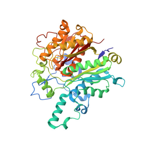
    FabF (3-oxoacyl-[acyl-carrier-protein] synthase 2), which catalyses the rate limiting condensation reaction in the fatty acid synthesis II pathway, is an attractive target for new antibiotics. Here, we focus on FabF from P. aeruginosa (PaFabF) as antibiotics against this pathogen are urgently needed. To facilitate exploration of this target we have set up an experimental toolbox consisting of binding assays using bio-layer interferometry (BLI) as well as saturation transfer difference (STD) and WaterLOGSY NMR in addition to robust conditions for structure determination. The suitability of the toolbox to support structure-based design of FabF inhibitors was demonstrated through the validation of hits obtained from virtual screening. Screening a library of almost 5 million compounds resulted in 6 compounds for which binding into the malonyl-binding site of FabF was shown. For one of the hits, the crystal structure in complex with PaFabF was determined. Based on the obtained binding mode, analogues were designed and synthesised, but affinity could not be improved. This work has laid the foundation for structure-based exploration of PaFabF.


  • Organizational Affiliation
    • Department of Biomedicine, University of Bergen, Jonas Lies Vei 91, 5020, Bergen, Norway.

Macromolecule Content 

  • Total Structure Weight: 88.52 kDa 
  • Atom Count: 6,725 
  • Modeled Residue Count: 824 
  • Deposited Residue Count: 824 
  • Unique protein chains: 1

Macromolecules

Find similar proteins by:|  3D Structure
Entity ID: 1
MoleculeChains  Sequence LengthOrganismDetailsImage
3-oxoacyl-[acyl-carrier-protein] synthase 2
A, B
412Pseudomonas aeruginosaMutation(s): 1 
EC: 2.3.1.179
UniProt
Find proteins for O54440 (Pseudomonas aeruginosa)
Explore O54440 
Go to UniProtKB:  O54440
Entity Groups
Sequence Clusters30% Identity50% Identity70% Identity90% Identity95% Identity100% Identity
UniProt GroupO54440
Sequence Annotations
Expand
Reference Sequence

Small Molecules

Ligands 4 Unique
IDChains Name / Formula / InChI Key2D Diagram3D Interactions
PMN

Query on PMN


Download:Ideal Coordinates CCD File
C [auth A],
J [auth B]
PLATENSIMYCIN
C24 H27 N O7
CSOMAHTTWTVBFL-OFBLZTNGSA-N
GOL

Query on GOL


Download:Ideal Coordinates CCD File
Q [auth B]GLYCEROL
C3 H8 O3
PEDCQBHIVMGVHV-UHFFFAOYSA-N
DMS

Query on DMS


Download:Ideal Coordinates CCD File
D [auth A]
E [auth A]
F [auth A]
G [auth A]
H [auth A]
D [auth A],
E [auth A],
F [auth A],
G [auth A],
H [auth A],
K [auth B],
L [auth B],
M [auth B],
N [auth B],
O [auth B],
P [auth B]
DIMETHYL SULFOXIDE
C2 H6 O S
IAZDPXIOMUYVGZ-UHFFFAOYSA-N
K

Query on K


Download:Ideal Coordinates CCD File
I [auth A]POTASSIUM ION
K
NPYPAHLBTDXSSS-UHFFFAOYSA-N

Experimental Data & Validation

Experimental Data

  • Method: X-RAY DIFFRACTION
  • Resolution: 1.80 Å
  • R-Value Free:  0.188 (Depositor), 0.195 (DCC) 
  • R-Value Work:  0.157 (Depositor) 
  • R-Value Observed: 0.159 (Depositor) 
Space Group: P 21 21 21
Unit Cell:
Length ( Å )Angle ( ˚ )
a = 84.024α = 90
b = 137.199β = 90
c = 65.692γ = 90
Software Package:
Software NamePurpose
REFMACrefinement
xia2data reduction
XSCALEdata scaling

Structure Validation

View Full Validation Report


Ligand Structure Quality Assessment 


Entry History 

& Funding Information

Deposition Data


Funding OrganizationLocationGrant Number
Research Council of NorwayNorway273588

Revision History  (Full details and data files)

  • Version 1.0: 2021-08-25
    Type: Initial release
  • Version 1.1: 2021-09-22
    Changes: Data collection, Database references
  • Version 1.2: 2024-01-31
    Changes: Data collection, Refinement description
  • Version 1.3: 2024-11-06
    Changes: Structure summary