7O6E | pdb_00007o6e

2.12 A cryo-EM structure of Mycobacterium tuberculosis Ferritin


Experimental Data Snapshot

  • Method: ELECTRON MICROSCOPY
  • Resolution: 2.10 Å
  • Aggregation State: PARTICLE 
  • Reconstruction Method: SINGLE PARTICLE 

Starting Model: experimental
View more details

wwPDB Validation 3D Report Full Report

Validation slider image for 7O6E

This is version 1.2 of the entry. See complete history

Literature

Mycobacterium tuberculosis ferritin: a suitable workhorse protein for cryo-EM development.

Gijsbers, A.Zhang, Y.Gao, Y.Peters, P.J.Ravelli, R.B.G.

(2021) Acta Crystallogr D Struct Biol 77: 1077-1083

  • DOI: https://doi.org/10.1107/S2059798321007233
  • Primary Citation Related Structures: 
    7O6E

  • PubMed Abstract: 

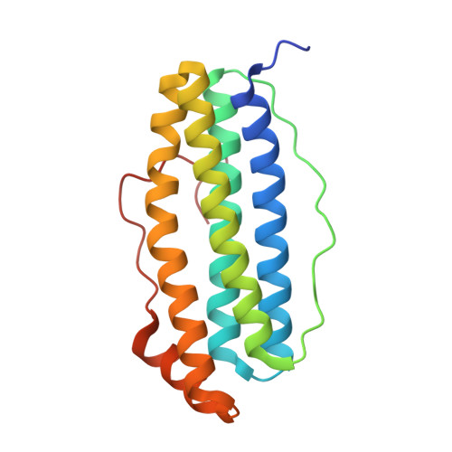
    The use of cryo-EM continues to expand worldwide and calls for good-quality standard proteins with simple protocols for their production. Here, a straightforward expression and purification protocol is presented that provides an apoferritin, bacterioferritin B (BfrB), from Mycobacterium tuberculosis with high yield and purity. A 2.12 Å resolution cryo-EM structure of BfrB is reported, showing the typical cage-like oligomer constituting of 24 monomers related by 432 symmetry. However, it also contains a unique C-terminal extension (164-181), which loops into the cage region of the shell and provides extra stability to the protein. Part of this region was ambiguous in previous crystal structures but could be built within the cryo-EM map. These findings and this protocol could serve the growing cryo-EM community in characterizing and pushing the limits of their electron microscopes and workflows.


  • Organizational Affiliation
    • Maastricht Multimodal Molecular Imaging Institute, Division of Nanoscopy, Maastricht University, Universiteitssingel 50, 6229 ER Maastricht, The Netherlands.

Macromolecule Content 

  • Total Structure Weight: 491.13 kDa 
  • Atom Count: 35,304 
  • Modeled Residue Count: 4,248 
  • Deposited Residue Count: 4,344 
  • Unique protein chains: 1

Macromolecules

Find similar proteins by:|  3D Structure
Entity ID: 1
MoleculeChains  Sequence LengthOrganismDetailsImage
Ferritin BfrB181Mycobacterium tuberculosis H37RvMutation(s): 0 
Gene Names: bfrB
EC: 1.16.3.1
UniProt
Find proteins for P9WNE5 (Mycobacterium tuberculosis (strain ATCC 25618 / H37Rv))
Explore P9WNE5 
Go to UniProtKB:  P9WNE5
Entity Groups
Sequence Clusters30% Identity50% Identity70% Identity90% Identity95% Identity100% Identity
UniProt GroupP9WNE5
Sequence Annotations
Expand
Reference Sequence

Experimental Data & Validation

Experimental Data

  • Method: ELECTRON MICROSCOPY
  • Resolution: 2.10 Å
  • Aggregation State: PARTICLE 
  • Reconstruction Method: SINGLE PARTICLE 
EM Software:
TaskSoftware PackageVersion
MODEL REFINEMENTPHENIX1.18.2
RECONSTRUCTIONRELION3.1

Structure Validation

View Full Validation Report


Entry History 

& Funding Information

Deposition Data


Funding OrganizationLocationGrant Number
European Union (EU)European Union766970
Netherlands Organisation for Scientific Research (NWO)Netherlands184.034.014

Revision History  (Full details and data files)

  • Version 1.0: 2021-05-19
    Type: Initial release
  • Version 1.1: 2021-09-15
    Changes: Database references
  • Version 1.2: 2024-07-10
    Changes: Data collection, Refinement description