7O5V | pdb_00007o5v

Crystal structure of holo-H44A mutant of Hydroxy ketone aldolase (SwHKA) from Sphingomonas wittichii RW1, in complex with Hydroxypyruvate


Experimental Data Snapshot

  • Method: X-RAY DIFFRACTION
  • Resolution: 1.95 Å
  • R-Value Free: 
    0.212 (Depositor), 0.213 (DCC) 
  • R-Value Work: 
    0.162 (Depositor), 0.162 (DCC) 

Starting Model: experimental
View more details

wwPDB Validation 3D Report Full Report

Validation slider image for 7O5V

Ligand Structure Quality Assessment 


This is version 1.3 of the entry. See complete history

Literature

Substrate Induced Movement of the Metal Cofactor between Active and Resting State.

Marsden, S.R.Wijma, H.J.Mohr, M.K.F.Justo, I.Hagedoorn, P.L.Laustsen, J.Jeffries, C.M.Svergun, D.Mestrom, L.McMillan, D.G.G.Bento, I.Hanefeld, U.

(2022) Angew Chem Int Ed Engl 61: e202213338-e202213338

  • DOI: https://doi.org/10.1002/anie.202213338
  • Primary Citation Related Structures: 
    7NNK, 7NR1, 7NUJ, 7O5I, 7O5R, 7O5V, 7O5W, 7O87, 7O9R, 7OBU, 8ADQ

  • PubMed Abstract: 

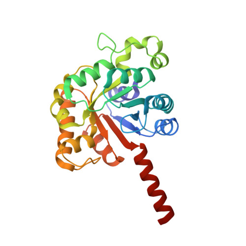
    Regulation of enzyme activity is vital for living organisms. In metalloenzymes, far-reaching rearrangements of the protein scaffold are generally required to tune the metal cofactor's properties by allosteric regulation. Here structural analysis of hydroxyketoacid aldolase from Sphingomonas wittichii RW1 (SwHKA) revealed a dynamic movement of the metal cofactor between two coordination spheres without protein scaffold rearrangements. In its resting state configuration (M 2+ R ), the metal constitutes an integral part of the dimer interface within the overall hexameric assembly, but sterical constraints do not allow for substrate binding. Conversely, a second coordination sphere constitutes the catalytically active state (M 2+ A ) at 2.4 Å distance. Bidentate coordination of a ketoacid substrate to M 2+ A affords the overall lowest energy complex, which drives the transition from M 2+ R to M 2+ A . While not described earlier, this type of regulation may be widespread and largely overlooked due to low occupancy of some of its states in protein crystal structures.


  • Organizational Affiliation
    • Biokatalyse, Afdeling Biotechnologie, Technische Universiteit Delft, van der Maasweg 9, 2629HZ, Delft, The Netherlands.

Macromolecule Content 

  • Total Structure Weight: 53.55 kDa 
  • Atom Count: 3,966 
  • Modeled Residue Count: 501 
  • Deposited Residue Count: 502 
  • Unique protein chains: 1

Macromolecules

Find similar proteins by:|  3D Structure
Entity ID: 1
MoleculeChains  Sequence LengthOrganismDetailsImage
HpcH/HpaI aldolaseA [auth AAA],
B [auth BBB]
251Rhizorhabdus wittichii RW1Mutation(s): 1 
Gene Names: Swit_5035

Small Molecules

Ligands 4 Unique
IDChains Name / Formula / InChI Key2D Diagram3D Interactions
3PY
(Subject of Investigation/LOI)

Query on 3PY


Download:Ideal Coordinates CCD File
F [auth AAA]3-HYDROXYPYRUVIC ACID
C3 H4 O4
HHDDCCUIIUWNGJ-UHFFFAOYSA-N
BR

Query on BR


Download:Ideal Coordinates CCD File
D [auth AAA],
G [auth AAA]
BROMIDE ION
Br
CPELXLSAUQHCOX-UHFFFAOYSA-M
K

Query on K


Download:Ideal Coordinates CCD File
E [auth AAA],
I [auth BBB]
POTASSIUM ION
K
NPYPAHLBTDXSSS-UHFFFAOYSA-N
MG
(Subject of Investigation/LOI)

Query on MG


Download:Ideal Coordinates CCD File
C [auth AAA],
H [auth BBB]
MAGNESIUM ION
Mg
JLVVSXFLKOJNIY-UHFFFAOYSA-N

Experimental Data & Validation

Experimental Data

  • Method: X-RAY DIFFRACTION
  • Resolution: 1.95 Å
  • R-Value Free:  0.212 (Depositor), 0.213 (DCC) 
  • R-Value Work:  0.162 (Depositor), 0.162 (DCC) 
Space Group: H 3
Unit Cell:
Length ( Å )Angle ( ˚ )
a = 70.914α = 90
b = 70.914β = 90
c = 222.572γ = 120
Software Package:
Software NamePurpose
REFMACrefinement
Aimlessdata scaling
XDSdata reduction
MOLREPphasing

Structure Validation

View Full Validation Report


Ligand Structure Quality Assessment 


Entry History 

Deposition Data

Revision History  (Full details and data files)

  • Version 1.0: 2022-11-16
    Type: Initial release
  • Version 1.1: 2022-11-23
    Changes: Database references
  • Version 1.2: 2022-12-07
    Changes: Database references
  • Version 1.3: 2024-01-31
    Changes: Data collection, Refinement description