7O3B | pdb_00007o3b

Crystal structure of the TTBK2-CEP164 complex bound to a camelid nanobody


Experimental Data Snapshot

  • Method: X-RAY DIFFRACTION
  • Resolution: 2.40 Å
  • R-Value Free: 
    0.239 (Depositor), 0.251 (DCC) 
  • R-Value Work: 
    0.189 (Depositor) 
  • R-Value Observed: 
    0.192 (Depositor) 

Starting Model: experimental
View more details

wwPDB Validation 3D Report Full Report

Validation slider image for 7O3B

This is version 1.4 of the entry. See complete history

Literature

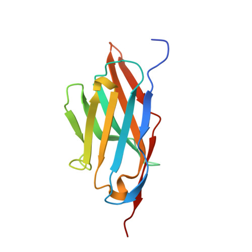
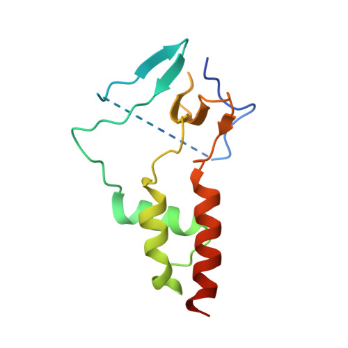
Molecular mechanisms underlying the role of the centriolar CEP164-TTBK2 complex in ciliopathies.

Rosa E Silva, I.Bino, L.Johnson, C.M.Rutherford, T.J.Neuhaus, D.Andreeva, A.Cajanek, L.van Breugel, M.

(2022) Structure 30: 114-128.e9

  • DOI: https://doi.org/10.1016/j.str.2021.08.007
  • Primary Citation Related Structures: 
    7NWJ, 7O06, 7O0S, 7O3B

  • PubMed Abstract: 

    Cilia formation is essential for human life. One of the earliest events in the ciliogenesis program is the recruitment of tau-tubulin kinase 2 (TTBK2) by the centriole distal appendage component CEP164. Due to the lack of high-resolution structural information on this complex, it is unclear how it is affected in human ciliopathies such as nephronophthisis. Furthermore, it is poorly understood if binding to CEP164 influences TTBK2 activities. Here, we present a detailed biochemical, structural, and functional analysis of the CEP164-TTBK2 complex and demonstrate how it is compromised by two ciliopathic mutations in CEP164. Moreover, we also provide insights into how binding to CEP164 is coordinated with TTBK2 activities. Together, our data deepen our understanding of a crucial step in cilia formation and will inform future studies aimed at restoring CEP164 functionality in a debilitating human ciliopathy.


  • Organizational Affiliation
    • Queen Mary University of London, School of Biological and Chemical Sciences, 2 Newark Street, London E1 2AT, UK; Medical Research Council - Laboratory of Molecular Biology, Francis Crick Avenue, Cambridge CB2 0QH, UK. Electronic address: ivan.silva@alumni.usp.br.

Macromolecule Content 

  • Total Structure Weight: 84.45 kDa 
  • Atom Count: 5,678 
  • Modeled Residue Count: 709 
  • Deposited Residue Count: 783 
  • Unique protein chains: 2

Macromolecules

Find similar proteins by:|  3D Structure
Entity ID: 1
MoleculeChains  Sequence LengthOrganismDetailsImage
Nanobody 36Z
A, B, C
126Camelidae mixed libraryMutation(s): 0 
Find similar proteins by:|  3D Structure
Entity ID: 2
MoleculeChains  Sequence LengthOrganismDetailsImage
Tau-tubulin kinase 2,Centrosomal protein of 164 kDaD [auth G],
E [auth H],
F [auth I]
135Homo sapiensMutation(s): 0 
Gene Names: TTBK2KIAA0847CEP164KIAA1052NPHP15
EC: 2.7.11.1
UniProt & NIH Common Fund Data Resources
Find proteins for Q6IQ55 (Homo sapiens)
Explore Q6IQ55 
Go to UniProtKB:  Q6IQ55
PHAROS:  Q6IQ55
GTEx:  ENSG00000128881 
Find proteins for Q9UPV0 (Homo sapiens)
Explore Q9UPV0 
Go to UniProtKB:  Q9UPV0
PHAROS:  Q9UPV0
GTEx:  ENSG00000110274 
Entity Groups
Sequence Clusters30% Identity50% Identity70% Identity90% Identity95% Identity100% Identity
UniProt GroupsQ9UPV0Q6IQ55
Sequence Annotations
Expand
Reference Sequence

Experimental Data & Validation

Experimental Data

  • Method: X-RAY DIFFRACTION
  • Resolution: 2.40 Å
  • R-Value Free:  0.239 (Depositor), 0.251 (DCC) 
  • R-Value Work:  0.189 (Depositor) 
  • R-Value Observed: 0.192 (Depositor) 
Space Group: C 2 2 21
Unit Cell:
Length ( Å )Angle ( ˚ )
a = 70.615α = 90
b = 127.741β = 90
c = 218.881γ = 90
Software Package:
Software NamePurpose
REFMACrefinement
PHENIXrefinement
DIALSdata reduction
Aimlessdata scaling
PHASERphasing

Structure Validation

View Full Validation Report


Entry History 

& Funding Information

Deposition Data


Funding OrganizationLocationGrant Number
Medical Research Council (MRC, United Kingdom)United KingdomMC_UP_1201/3

Revision History  (Full details and data files)

  • Version 1.0: 2021-09-15
    Type: Initial release
  • Version 1.1: 2021-09-22
    Changes: Data collection, Database references
  • Version 1.2: 2022-01-19
    Changes: Data collection, Database references
  • Version 1.3: 2022-12-21
    Changes: Database references
  • Version 1.4: 2024-01-31
    Changes: Data collection, Refinement description