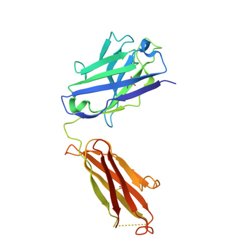
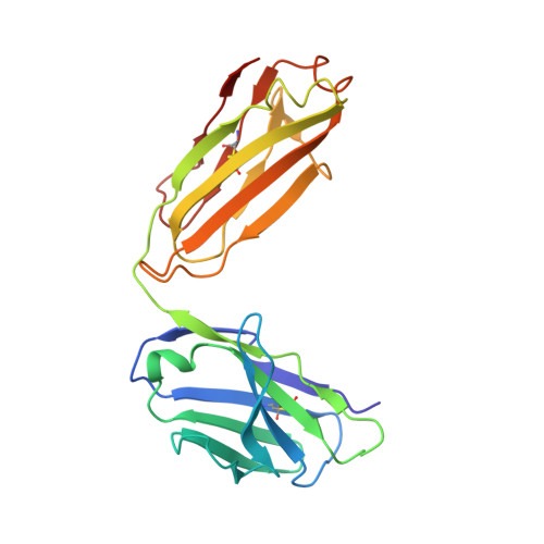
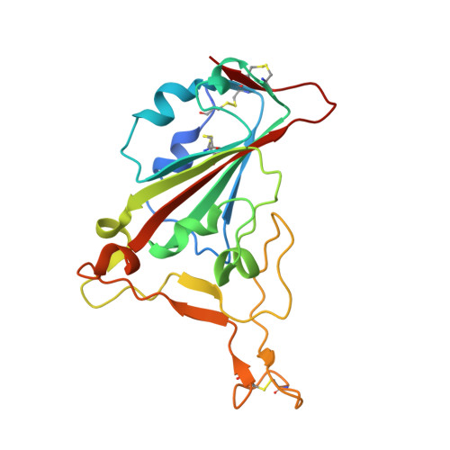
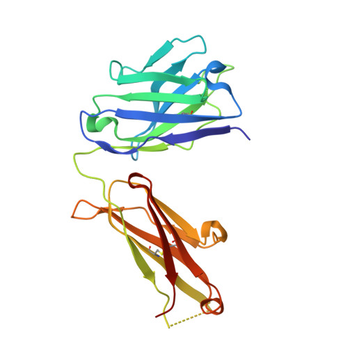
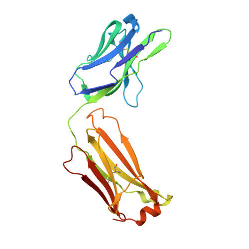
Terminating the SARS-CoV-2 pandemic relies upon pan-global vaccination. Current vaccines elicit neutralizing antibody responses to the virus spike derived from early isolates. However, new strains have emerged with multiple mutations, including P.1 from Brazil, B.1.351 from South Africa, and B.1.1.7 from the UK (12, 10, and 9 changes in the spike, respectively). All have mutations in the ACE2 binding site, with P.1 and B.1.351 having a virtually identical triplet (E484K, K417N/T, and N501Y), which we show confer similar increased affinity for ACE2. We show that, surprisingly, P.1 is significantly less resistant to naturally acquired or vaccine-induced antibody responses than B.1.351, suggesting that changes outside the receptor-binding domain (RBD) impact neutralization. Monoclonal antibody (mAb) 222 neutralizes all three variants despite interacting with two of the ACE2-binding site mutations. We explain this through structural analysis and use the 222 light chain to largely restore neutralization potency to a major class of public antibodies.
Organizational Affiliation:
Wellcome Centre for Human Genetics, Nuffield Department of Medicine, University of Oxford, Oxford, UK. Electronic address: dwanwisa@well.ox.ac.uk.
Division of Structural Biology, Nuffield Department of Medicine, University of Oxford, The Wellcome Centre for Human Genetics, Oxford, UK.
Wellcome Centre for Human Genetics, Nuffield Department of Medicine, University of Oxford, Oxford, UK.
Wellcome Centre for Human Genetics, Nuffield Department of Medicine, University of Oxford, Oxford, UK; Chinese Academy of Medical Science (CAMS) Oxford Institute (COI), University of Oxford, Oxford, UK.
Wellcome Centre for Human Genetics, Nuffield Department of Medicine, University of Oxford, Oxford, UK; Oxford University Hospitals NHS Foundation Trust, Oxford, UK.
Oxford University Hospitals NHS Foundation Trust, Oxford, UK; Peter Medawar Building for Pathogen Research, Oxford, UK; Nuffield Department of Clinical Neurosciences, University of Oxford, Oxford, UK.
Institute of Global Health, University of Siena, Siena, Brazil; Department of Paediatrics, University of Oxford, Oxford, UK.
Laboratório de Ecologia de Doenças Transmissíveis na Amazônia, Instituto Leônidas e Maria Deane, FIOCRUZ, Manaus, Amazonas, Brazil.
Fundação de Vigilância em Saúde do Amazonas, Manaus, Amazonas, Brazil.
Laboratory of Respiratory Viruses and Measles, Oswaldo Cruz Institute, FIOCRUZ, Rio de Janeiro, Brazil.
Laboratory of Respiratory Viruses and Measles, Oswaldo Cruz Institute, FIOCRUZ, Rio de Janeiro, Brazil; Department of Veterinary Integrative Biosciences, Texas A&M University, College Station, TX, United States.
NIHR Oxford Biomedical Research Centre, Oxford, UK; Oxford Vaccine Group, Department of Paediatrics, University of Oxford, Oxford, UK.
Worthing Hospital, Worthing, UK.
Chinese Academy of Medical Science (CAMS) Oxford Institute (COI), University of Oxford, Oxford, UK; MRC Human Immunology Unit, MRC Weatherall Institute of Molecular Medicine, Radcliffe Department of Medicine, University of Oxford, Oxford, UK; Nuffield Department of Medicine, University of Oxford, Oxford, UK.
Wellcome Centre for Human Genetics, Nuffield Department of Medicine, University of Oxford, Oxford, UK; Chinese Academy of Medical Science (CAMS) Oxford Institute (COI), University of Oxford, Oxford, UK; Nuffield Department of Medicine, University of Oxford, Oxford, UK.
Nuffield Department of Medicine, University of Oxford, Oxford, UK.
Jenner Institute, Nuffield Department of Medicine, University of Oxford, Oxford, UK.
Wellcome Centre for Human Genetics, Nuffield Department of Medicine, University of Oxford, Oxford, UK; National Infection Service, Public Health England (PHE), Porton Down, Salisbury, UK.
Oxford University Hospitals NHS Foundation Trust, Oxford, UK; Peter Medawar Building for Pathogen Research, Oxford, UK; NIHR Oxford Biomedical Research Centre, Oxford, UK; Translational Gastroenterology Unit, University of Oxford, Oxford, UK.
Oxford University Hospitals NHS Foundation Trust, Oxford, UK; Peter Medawar Building for Pathogen Research, Oxford, UK; Centre For Tropical Medicine and Global Health, Nuffield Department of Medicine, University of Oxford, Oxford, UK; Mahidol-Oxford Tropical Medicine Research Unit, Bangkok, Thailand, Department of Medicine, University of Oxford, Oxford, UK.
Wellcome Centre for Human Genetics, Nuffield Department of Medicine, University of Oxford, Oxford, UK; Chinese Academy of Medical Science (CAMS) Oxford Institute (COI), University of Oxford, Oxford, UK; Siriraj Center of Research Excellence in Dengue & Emerging Pathogens, Dean Office for Research, Faculty of Medicine Siriraj Hospital, Mahidol University, Thailand. Electronic address: jmongkol@well.ox.ac.uk.
Division of Structural Biology, Nuffield Department of Medicine, University of Oxford, The Wellcome Centre for Human Genetics, Oxford, UK. Electronic address: ren@strubi.ox.ac.uk.
Wellcome Centre for Human Genetics, Nuffield Department of Medicine, University of Oxford, Oxford, UK; Division of Structural Biology, Nuffield Department of Medicine, University of Oxford, The Wellcome Centre for Human Genetics, Oxford, UK; Chinese Academy of Medical Science (CAMS) Oxford Institute (COI), University of Oxford, Oxford, UK; Diamond Light Source Ltd, Harwell Science & Innovation Campus, Didcot, UK; Instruct-ERIC, Oxford House, Parkway Court, John Smith Drive, Oxford, UK. Electronic address: dave@strubi.ox.ac.uk.
Wellcome Centre for Human Genetics, Nuffield Department of Medicine, University of Oxford, Oxford, UK; Chinese Academy of Medical Science (CAMS) Oxford Institute (COI), University of Oxford, Oxford, UK. Electronic address: gavin.screaton@medsci.ox.ac.uk.