7NTK | pdb_00007ntk

PALS1 PDZ1 domain with SARS-CoV-2_E PBM complex


Experimental Data Snapshot

  • Method: X-RAY DIFFRACTION
  • Resolution: 1.90 Å
  • R-Value Free: 
    0.229 (Depositor), 0.230 (DCC) 
  • R-Value Work: 
    0.196 (Depositor), 0.197 (DCC) 
  • R-Value Observed: 
    0.198 (Depositor) 

Starting Model: experimental
View more details

wwPDB Validation 3D Report Full Report

Validation slider image for 7NTK

This is version 1.2 of the entry. See complete history

Literature

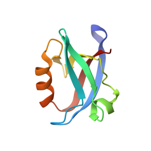
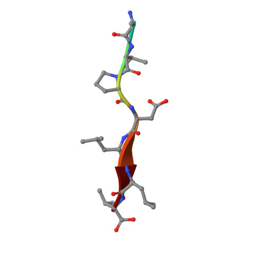
Structural basis of coronavirus E protein interactions with human PALS1 PDZ domain.

Javorsky, A.Humbert, P.O.Kvansakul, M.

(2021) Commun Biol 4: 724-724

  • DOI: https://doi.org/10.1038/s42003-021-02250-7
  • Primary Citation Related Structures: 
    7NTJ, 7NTK

  • PubMed Abstract: 

    SARS-CoV-2 infection leads to coronavirus disease 2019 (COVID-19), which is associated with severe and life-threatening pneumonia and respiratory failure. However, the molecular basis of these symptoms remains unclear. SARS-CoV-1 E protein interferes with control of cell polarity and cell-cell junction integrity in human epithelial cells by binding to the PALS1 PDZ domain, a key component of the Crumbs polarity complex. We show that C-terminal PDZ binding motifs of SARS-CoV-1 and SARS-CoV-2 E proteins bind the PALS1 PDZ domain with 29.6 and 22.8 μM affinity, whereas the related sequence from MERS-CoV did not bind. We then determined crystal structures of PALS1 PDZ domain bound to both SARS-CoV-1 and SARS-CoV-2 E protein PDZ binding motifs. Our findings establish the structural basis for SARS-CoV-1/2 mediated subversion of Crumbs polarity signalling and serve as a platform for the development of small molecule inhibitors to suppress SARS-CoV-1/2 mediated disruption of polarity signalling in epithelial cells.


  • Organizational Affiliation
    • Department of Biochemistry & Genetics, La Trobe Institute for Molecular Science, La Trobe University, Melbourne, Vic, Australia.

Macromolecule Content 

  • Total Structure Weight: 41.66 kDa 
  • Atom Count: 3,155 
  • Modeled Residue Count: 370 
  • Deposited Residue Count: 380 
  • Unique protein chains: 2

Macromolecules

Find similar proteins by:|  3D Structure
Entity ID: 1
MoleculeChains  Sequence LengthOrganismDetailsImage
MAGUK p55 subfamily member 5
A, B, D, F
87Homo sapiensMutation(s): 0 
Gene Names: MPP5PALS1
UniProt & NIH Common Fund Data Resources
Find proteins for Q8N3R9 (Homo sapiens)
Explore Q8N3R9 
Go to UniProtKB:  Q8N3R9
PHAROS:  Q8N3R9
GTEx:  ENSG00000072415 
Entity Groups
Sequence Clusters30% Identity50% Identity70% Identity90% Identity95% Identity100% Identity
UniProt GroupQ8N3R9
Sequence Annotations
Expand
Reference Sequence
Find similar proteins by:  Sequence   |   3D Structure  
Entity ID: 2
MoleculeChains  Sequence LengthOrganismDetailsImage
Envelope small membrane protein
C, E, G, H
8Severe acute respiratory syndrome coronavirus 2Mutation(s): 0 
UniProt
Find proteins for P0DTC4 (Severe acute respiratory syndrome coronavirus 2)
Explore P0DTC4 
Go to UniProtKB:  P0DTC4
Entity Groups
UniProt GroupP0DTC4
Sequence Annotations
Expand
Reference Sequence

Experimental Data & Validation

Experimental Data

  • Method: X-RAY DIFFRACTION
  • Resolution: 1.90 Å
  • R-Value Free:  0.229 (Depositor), 0.230 (DCC) 
  • R-Value Work:  0.196 (Depositor), 0.197 (DCC) 
  • R-Value Observed: 0.198 (Depositor) 
Space Group: P 1 21 1
Unit Cell:
Length ( Å )Angle ( ˚ )
a = 28.688α = 90
b = 103.762β = 103.979
c = 59.076γ = 90
Software Package:
Software NamePurpose
PHENIXrefinement
DIALSdata reduction
Aimlessdata scaling
PHASERphasing

Structure Validation

View Full Validation Report


Entry History 

Deposition Data

Revision History  (Full details and data files)

  • Version 1.0: 2021-06-16
    Type: Initial release
  • Version 1.1: 2021-07-21
    Changes: Database references
  • Version 1.2: 2024-01-31
    Changes: Data collection, Database references, Refinement description