7NG0 | pdb_00007ng0

Crystal structure of N- and C-terminally truncated Geobacillus thermoleovorans nucleoid occlusion protein Noc


Experimental Data Snapshot

  • Method: X-RAY DIFFRACTION
  • Resolution: 2.95 Å
  • R-Value Free: 
    0.288 (Depositor), 0.289 (DCC) 
  • R-Value Work: 
    0.267 (Depositor), 0.269 (DCC) 
  • R-Value Observed: 
    0.269 (Depositor) 

Starting Model: experimental
View more details

wwPDB Validation 3D Report Full Report

Validation slider image for 7NG0

This is version 1.3 of the entry. See complete history

Literature

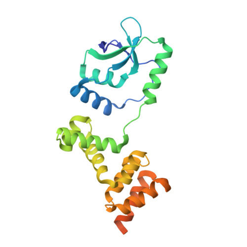
CTP regulates membrane-binding activity of the nucleoid occlusion protein Noc.

Jalal, A.S.B.Tran, N.T.Wu, L.J.Ramakrishnan, K.Rejzek, M.Gobbato, G.Stevenson, C.E.M.Lawson, D.M.Errington, J.Le, T.B.K.

(2021) Mol Cell 81: 3623-3636.e6

  • DOI: https://doi.org/10.1016/j.molcel.2021.06.025
  • Primary Citation Related Structures: 
    7NFU, 7NG0

  • PubMed Abstract: 

    ATP- and GTP-dependent molecular switches are extensively used to control functions of proteins in a wide range of biological processes. However, CTP switches are rarely reported. Here, we report that a nucleoid occlusion protein Noc is a CTPase enzyme whose membrane-binding activity is directly regulated by a CTP switch. In Bacillus subtilis, Noc nucleates on 16 bp NBS sites before associating with neighboring non-specific DNA to form large membrane-associated nucleoprotein complexes to physically occlude assembly of the cell division machinery. By in vitro reconstitution, we show that (1) CTP is required for Noc to form the NBS-dependent nucleoprotein complex, and (2) CTP binding, but not hydrolysis, switches Noc to a membrane-active state. Overall, we suggest that CTP couples membrane-binding activity of Noc to nucleoprotein complex formation to ensure productive recruitment of DNA to the bacterial cell membrane for nucleoid occlusion activity.


  • Organizational Affiliation
    • Department of Molecular Microbiology, John Innes Centre, Norwich, NR4 7UH, UK.

Macromolecule Content 

  • Total Structure Weight: 26.49 kDa 
  • Atom Count: 1,520 
  • Modeled Residue Count: 199 
  • Deposited Residue Count: 228 
  • Unique protein chains: 1

Macromolecules

Find similar proteins by:|  3D Structure
Entity ID: 1
MoleculeChains  Sequence LengthOrganismDetailsImage
Nucleoid occlusion protein228Geobacillus thermoleovorans CCB_US3_UF5Mutation(s): 0 
Gene Names: nocGTCCBUS3UF5_39100

Small Molecules

Ligands 1 Unique
IDChains Name / Formula / InChI Key2D Diagram3D Interactions
SO4

Query on SO4


Download:Ideal Coordinates CCD File
B [auth A]SULFATE ION
O4 S
QAOWNCQODCNURD-UHFFFAOYSA-L

Experimental Data & Validation

Experimental Data

  • Method: X-RAY DIFFRACTION
  • Resolution: 2.95 Å
  • R-Value Free:  0.288 (Depositor), 0.289 (DCC) 
  • R-Value Work:  0.267 (Depositor), 0.269 (DCC) 
  • R-Value Observed: 0.269 (Depositor) 
Space Group: C 2 2 21
Unit Cell:
Length ( Å )Angle ( ˚ )
a = 105.067α = 90
b = 106.562β = 90
c = 42.215γ = 90
Software Package:
Software NamePurpose
Aimlessdata scaling
REFMACrefinement
PDB_EXTRACTdata extraction
DIALSdata reduction
PHASERphasing

Structure Validation

View Full Validation Report


Entry History 

& Funding Information

Deposition Data


Funding OrganizationLocationGrant Number
Royal SocietyUnited KingdomURF-R-201020
Wellcome TrustUnited Kingdom209500
Royal SocietyUnited KingdomRG150448
Biotechnology and Biological Sciences Research Council (BBSRC)United KingdomBBS-E-J-000C0683

Revision History  (Full details and data files)

  • Version 1.0: 2021-02-17
    Type: Initial release
  • Version 1.1: 2021-07-28
    Changes: Database references
  • Version 1.2: 2021-09-15
    Changes: Database references
  • Version 1.3: 2024-01-31
    Changes: Data collection, Refinement description