


Uncovering a novel mechanism of enzyme activation in multimeric azoreductases
Hitchings, R., Ryan, A., Arcinas, A.J., Ghosh, A., Almo, S.C., Kelly, L.To be published.
Experimental Data Snapshot
Starting Model: experimental
View more details
Entity ID: 1 | |||||
|---|---|---|---|---|---|
| Molecule | Chains | Sequence Length | Organism | Details | Image |
| FMN dependent NADH:quinone oxidoreductase | 223 | Klebsiella pneumoniae subsp. pneumoniae MGH 78578 | Mutation(s): 0 Gene Names: acpD, azoR, KPN_02994 EC: 1.6.5 (PDB Primary Data), 1.7.1.17 (PDB Primary Data) | 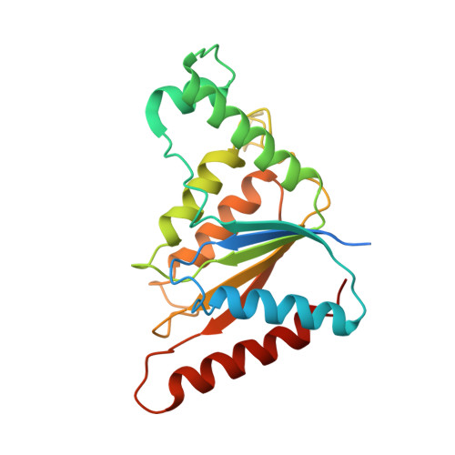 | |
UniProt | |||||
Entity Groups | |||||
| Sequence Clusters | 30% Identity50% Identity70% Identity90% Identity95% Identity100% Identity | ||||
| UniProt Group | A6TCS9 | ||||
Sequence AnnotationsExpand | |||||
Reference Sequence | |||||
| Ligands 2 Unique | |||||
|---|---|---|---|---|---|
| ID | Chains | Name / Formula / InChI Key | 2D Diagram | 3D Interactions | |
| FMN Download:Ideal Coordinates CCD File | E [auth A], H [auth B], I [auth C], L [auth D] | FLAVIN MONONUCLEOTIDE C17 H21 N4 O9 P FVTCRASFADXXNN-SCRDCRAPSA-N |  | ||
| 05G Download:Ideal Coordinates CCD File | F [auth A], G [auth B], J [auth C], K [auth C] | 6-hydroxy-5-[(E)-(2-methoxy-5-methyl-4-sulfophenyl)diazenyl]naphthalene-2-sulfonic acid C18 H16 N2 O8 S2 UQWIHFJXDRNUDP-FMQUCBEESA-N |  | ||
| Length ( Å ) | Angle ( ˚ ) |
|---|---|
| a = 60.407 | α = 90 |
| b = 83.336 | β = 100.26 |
| c = 87.305 | γ = 90 |
| Software Name | Purpose |
|---|---|
| PHENIX | refinement |
| MOSFLM | data reduction |
| Aimless | data scaling |
| PDB_EXTRACT | data extraction |
| PHASER | phasing |


