7N0A | pdb_00007n0a

Structure of Human Leukaemia Inhibitory Factor with Fab MSC1


Experimental Data Snapshot

  • Method: X-RAY DIFFRACTION
  • Resolution: 3.10 Å
  • R-Value Free: 
    0.284 (Depositor), 0.280 (DCC) 
  • R-Value Work: 
    0.232 (Depositor), 0.232 (DCC) 
  • R-Value Observed: 
    0.234 (Depositor) 

Starting Model: experimental
View more details

wwPDB Validation 3D Report Full Report

Validation slider image for 7N0A

Ligand Structure Quality Assessment 


This is version 1.4 of the entry. See complete history

Literature

Therapeutic Targeting of LIF Overcomes Macrophage-mediated Immunosuppression of the Local Tumor Microenvironment.

Hallett, R.M.Bonfill-Teixidor, E.Iurlaro, R.Arias, A.Raman, S.Bayliss, P.Egorova, O.Neva-Alejo, A.McGray, A.R.Lau, E.Bosch, A.Beilschmidt, M.Maetzel, D.Fransson, J.Huber-Ruano, I.Anido, J.Julien, J.P.Giblin, P.Seoane, J.

(2023) Clin Cancer Res 29: 791-804

  • DOI: https://doi.org/10.1158/1078-0432.CCR-21-1888
  • Primary Citation Related Structures: 
    7N0A

  • PubMed Abstract: 

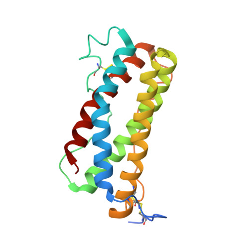
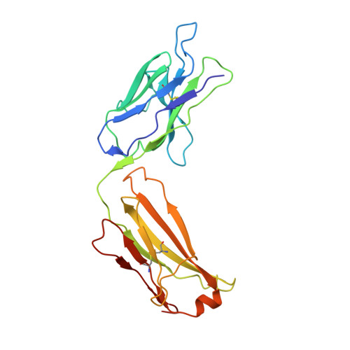
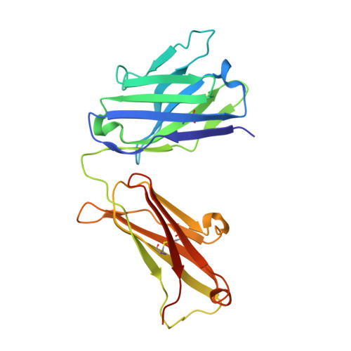
    Leukemia inhibitory factor (LIF) is a multifunctional cytokine with numerous reported roles in cancer and is thought to drive tumor development and progression. Characterization of LIF and clinical-stage LIF inhibitors would increase our understanding of LIF as a therapeutic target. We first tested the association of LIF expression with transcript signatures representing multiple processes regulating tumor development and progression. Next, we developed MSC-1, a high-affinity therapeutic antibody that potently inhibits LIF signaling and tested it in immune competent animal models of cancer. LIF was associated with signatures of tumor-associated macrophages (TAM) across 7,769 tumor samples spanning 22 solid tumor indications. In human tumors, LIF receptor was highly expressed within the macrophage compartment and LIF treatment drove macrophages to acquire immunosuppressive capacity. MSC-1 potently inhibited LIF signaling by binding an epitope that overlaps with the gp130 receptor binding site on LIF. MSC-1 showed monotherapy efficacy in vivo and drove TAMs to acquire antitumor and proinflammatory function in syngeneic colon cancer mouse models. Combining MSC-1 with anti-PD1 leads to strong antitumor response and a long-term tumor-free survival in a significant proportion of treated mice. Overall, our findings highlight LIF as a therapeutic target for cancer immunotherapy.


  • Organizational Affiliation
    • Northern Biologics, Toronto, Ontario, Canada.

Macromolecule Content 

  • Total Structure Weight: 69.32 kDa 
  • Atom Count: 4,755 
  • Modeled Residue Count: 611 
  • Deposited Residue Count: 625 
  • Unique protein chains: 3

Macromolecules

Find similar proteins by:|  3D Structure
Entity ID: 1
MoleculeChains  Sequence LengthOrganismDetailsImage
Leukemia inhibitory factorA [auth C]181Homo sapiensMutation(s): 0 
Gene Names: LIFHILDA
UniProt & NIH Common Fund Data Resources
Find proteins for P15018 (Homo sapiens)
Explore P15018 
Go to UniProtKB:  P15018
PHAROS:  P15018
GTEx:  ENSG00000128342 
Entity Groups
Sequence Clusters30% Identity50% Identity70% Identity90% Identity95% Identity100% Identity
UniProt GroupP15018
Glycosylation
Glycosylation Sites: 4Go to GlyGen: P15018-1
Sequence Annotations
Expand
Reference Sequence
Find similar proteins by:|  3D Structure
Entity ID: 2
MoleculeChains  Sequence LengthOrganismDetailsImage
MSC-1 Fab Light chainB [auth A]220Homo sapiensMutation(s): 0 
Find similar proteins by:|  3D Structure
Entity ID: 3
MoleculeChains  Sequence LengthOrganismDetailsImage
MSC-1 Fab Heavy chainC [auth B]224Homo sapiensMutation(s): 0 

Oligosaccharides

Help  
Entity ID: 4
MoleculeChains Length2D Diagram GlycosylationD Interactions
beta-D-mannopyranose-(1-4)-2-acetamido-2-deoxy-beta-D-glucopyranose-(1-4)-2-acetamido-2-deoxy-beta-D-glucopyranoseD [auth E]3N-Glycosylation
Glycosylation Resources
GlyTouCan: G15407YE
GlyCosmos: G15407YE
GlyGen: G15407YE

Experimental Data & Validation

Experimental Data

  • Method: X-RAY DIFFRACTION
  • Resolution: 3.10 Å
  • R-Value Free:  0.284 (Depositor), 0.280 (DCC) 
  • R-Value Work:  0.232 (Depositor), 0.232 (DCC) 
  • R-Value Observed: 0.234 (Depositor) 
Space Group: P 21 21 2
Unit Cell:
Length ( Å )Angle ( ˚ )
a = 60.572α = 90
b = 109.38β = 90
c = 115.691γ = 90
Software Package:
Software NamePurpose
PHENIXrefinement
XPREPdata reduction
XDSdata scaling
PHASERphasing

Structure Validation

View Full Validation Report


Ligand Structure Quality Assessment 


Entry History 

& Funding Information

Deposition Data


Funding OrganizationLocationGrant Number
Canada Research ChairsCanada950231604

Revision History  (Full details and data files)

  • Version 1.0: 2022-06-15
    Type: Initial release
  • Version 1.1: 2022-12-14
    Changes: Database references
  • Version 1.2: 2023-02-22
    Changes: Database references
  • Version 1.3: 2023-10-25
    Changes: Data collection, Refinement description
  • Version 1.4: 2024-10-09
    Changes: Structure summary