7MQX | pdb_00007mqx

P. putida mandelate racemase forms an oxobenzoxaborole adduct with 2-formylphenylboronic acid


Experimental Data Snapshot

  • Method: X-RAY DIFFRACTION
  • Resolution: 1.91 Å
  • R-Value Free: 
    0.215 (Depositor), 0.215 (DCC) 
  • R-Value Work: 
    0.187 (Depositor), 0.187 (DCC) 
  • R-Value Observed: 
    0.189 (Depositor) 

Starting Model: experimental
View more details

wwPDB Validation 3D Report Full Report

Validation slider image for 7MQX

Ligand Structure Quality Assessment 


This is version 1.3 of the entry. See complete history

Literature

Slow-Onset, Potent Inhibition of Mandelate Racemase by 2-Formylphenylboronic Acid. An Unexpected Adduct Clasps the Catalytic Machinery.

Douglas, C.D.Grandinetti, L.Easton, N.M.Kuehm, O.P.Hayden, J.A.Hamilton, M.C.St Maurice, M.Bearne, S.L.

(2021) Biochemistry 

  • DOI: https://doi.org/10.1021/acs.biochem.1c00374
  • Primary Citation Related Structures: 
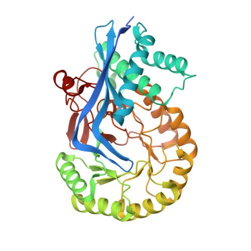
    7MQX

  • PubMed Abstract: 

    o -Carbonyl arylboronic acids such as 2-formylphenylboronic acid (2-FPBA) are employed in biocompatible conjugation reactions with the resulting iminoboronate adduct stabilized by an intramolecular N-B interaction. However, few studies have utilized these reagents as active site-directed enzyme inhibitors. We show that 2-FPBA is a potent reversible, slow-onset inhibitor of mandelate racemase (MR), an enzyme that has served as a valuable paradigm for understanding enzyme-catalyzed abstraction of an α-proton from a carbon acid substrate with a high p K a . Kinetic analysis of the progress curves for the slow onset of inhibition of wild-type MR using a two-step kinetic mechanism gave K i and K i * values of 5.1 ± 1.8 and 0.26 ± 0.08 μM, respectively. Hence, wild-type MR binds 2-FPBA with an affinity that exceeds that for the substrate by ∼3000-fold. K164R MR was inhibited by 2-FPBA, while K166R MR was not inhibited, indicating that Lys 166 was essential for inhibition. Unexpectedly, mass spectrometric analysis of the NaCNBH 3 -treated enzyme-inhibitor complex did not yield evidence of an iminoboronate adduct. 11 B nuclear magnetic resonance spectroscopy of the MR·2-FPBA complex indicated that the boron atom was sp 3 -hybridized (δ 6.0), consistent with dative bond formation. Surprisingly, X-ray crystallography revealed the formation of an N ζ -B dative bond between Lys 166 and 2-FPBA with intramolecular cyclization to form a benzoxaborole, rather than the expected iminoboronate. Thus, when o -carbonyl arylboronic acid reagents are employed to modify proteins, the structure of the resulting product depends on the protein architecture at the site of modification.


  • Organizational Affiliation
    • Department of Biochemistry and Molecular Biology, Dalhousie University, Halifax, NS B3H 4R2, Canada.

Macromolecule Content 

  • Total Structure Weight: 333.32 kDa 
  • Atom Count: 24,058 
  • Modeled Residue Count: 2,857 
  • Deposited Residue Count: 3,064 
  • Unique protein chains: 1

Macromolecules

Find similar proteins by:|  3D Structure
Entity ID: 1
MoleculeChains  Sequence LengthOrganismDetailsImage
Mandelate racemase
A, B, C, D, E
A, B, C, D, E, F, G, H
383Pseudomonas putidaMutation(s): 0 
Gene Names: mdlA
EC: 5.1.2.2
UniProt
Find proteins for P11444 (Pseudomonas putida)
Explore P11444 
Go to UniProtKB:  P11444
Entity Groups
Sequence Clusters30% Identity50% Identity70% Identity90% Identity95% Identity100% Identity
UniProt GroupP11444
Sequence Annotations
Expand
Reference Sequence

Small Molecules

Ligands 3 Unique
IDChains Name / Formula / InChI Key2D Diagram3D Interactions
ZMD
(Subject of Investigation/LOI)

Query on ZMD


Download:Ideal Coordinates CCD File
DA [auth E]
IA [auth F]
J [auth A]
NA [auth G]
O [auth B]
DA [auth E],
IA [auth F],
J [auth A],
NA [auth G],
O [auth B],
SA [auth H],
U [auth C],
Y [auth D]
(3S)-2,1-benzoxaborole-1,3(3H)-diol
C7 H7 B O3
GKXMUTRKNBAUGN-ZETCQYMHSA-N
EDO

Query on EDO


Download:Ideal Coordinates CCD File
AA [auth D]
BA [auth D]
EA [auth E]
FA [auth E]
GA [auth E]
AA [auth D],
BA [auth D],
EA [auth E],
FA [auth E],
GA [auth E],
JA [auth F],
K [auth A],
KA [auth F],
L [auth A],
LA [auth F],
M [auth A],
OA [auth G],
P [auth B],
PA [auth G],
Q [auth B],
QA [auth G],
R [auth B],
S [auth B],
TA [auth H],
UA [auth H],
V [auth C],
VA [auth H],
W [auth C],
Z [auth D]
1,2-ETHANEDIOL
C2 H6 O2
LYCAIKOWRPUZTN-UHFFFAOYSA-N
MG

Query on MG


Download:Ideal Coordinates CCD File
CA [auth E]
HA [auth F]
I [auth A]
MA [auth G]
N [auth B]
CA [auth E],
HA [auth F],
I [auth A],
MA [auth G],
N [auth B],
RA [auth H],
T [auth C],
X [auth D]
MAGNESIUM ION
Mg
JLVVSXFLKOJNIY-UHFFFAOYSA-N

Experimental Data & Validation

Experimental Data

  • Method: X-RAY DIFFRACTION
  • Resolution: 1.91 Å
  • R-Value Free:  0.215 (Depositor), 0.215 (DCC) 
  • R-Value Work:  0.187 (Depositor), 0.187 (DCC) 
  • R-Value Observed: 0.189 (Depositor) 
Space Group: C 1 2 1
Unit Cell:
Length ( Å )Angle ( ˚ )
a = 225.483α = 90
b = 149.43β = 129.34
c = 149.523γ = 90
Software Package:
Software NamePurpose
Aimlessdata scaling
PHASERphasing
PHENIXrefinement
PDB_EXTRACTdata extraction
XDSdata reduction

Structure Validation

View Full Validation Report


Ligand Structure Quality Assessment 


Entry History 

& Funding Information

Deposition Data


Funding OrganizationLocationGrant Number
Natural Sciences and Engineering Research Council (NSERC, Canada)CanadaRGPIN-2016-05083

Revision History  (Full details and data files)

  • Version 1.0: 2021-08-11
    Type: Initial release
  • Version 1.1: 2021-08-25
    Changes: Database references
  • Version 1.2: 2023-10-18
    Changes: Data collection, Refinement description
  • Version 1.3: 2024-10-23
    Changes: Structure summary