7MOW | pdb_00007mow

PTP1B F225I in complex with TCS401

  • Classification: HYDROLASE
  • Organism(s): Homo sapiens
  • Expression System: Escherichia coli
  • Mutation(s): Yes 

  • Deposited: 2021-05-03 Released: 2022-05-18 
  • Deposition Author(s): Torgeson, K.R., Page, R., Peti, W.
  • Funding Organization(s): American Diabetes Association, National Institutes of Health/National Institute of General Medical Sciences (NIH/NIGMS)

Experimental Data Snapshot

  • Method: X-RAY DIFFRACTION
  • Resolution: 1.80 Å
  • R-Value Free: 
    0.176 (Depositor), 0.167 (DCC) 
  • R-Value Work: 
    0.155 (Depositor) 
  • R-Value Observed: 
    0.156 (Depositor) 

Starting Model: experimental
View more details

wwPDB Validation 3D Report Full Report

Validation slider image for 7MOW

Ligand Structure Quality Assessment 


This is version 1.2 of the entry. See complete history

Literature

Conserved conformational dynamics determine enzyme activity.

Torgeson, K.R.Clarkson, M.W.Granata, D.Lindorff-Larsen, K.Page, R.Peti, W.

(2022) Sci Adv 8: eabo5546-eabo5546

  • DOI: https://doi.org/10.1126/sciadv.abo5546
  • Primary Citation Related Structures: 
    7MKZ, 7MN7, 7MN9, 7MNA, 7MNB, 7MNC, 7MND, 7MNE, 7MNF, 7MOU, 7MOV, 7MOW

  • PubMed Abstract: 

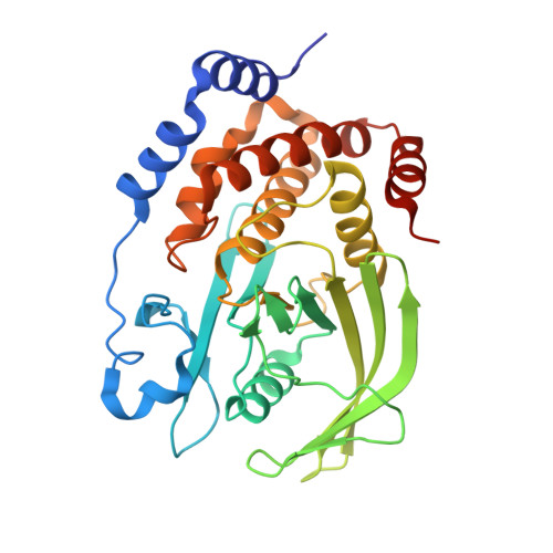
    Homologous enzymes often exhibit different catalytic rates despite a fully conserved active site. The canonical view is that an enzyme sequence defines its structure and function and, more recently, that intrinsic protein dynamics at different time scales enable and/or promote catalytic activity. Here, we show that, using the protein tyrosine phosphatase PTP1B, residues surrounding the PTP1B active site promote dynamically coordinated chemistry necessary for PTP1B function. However, residues distant to the active site also undergo distinct intermediate time scale dynamics and these dynamics are correlated with its catalytic activity and thus allow for different catalytic rates in this enzyme family. We identify these previously undetected motions using coevolutionary coupling analysis and nuclear magnetic resonance spectroscopy. Our findings strongly indicate that conserved dynamics drives the enzymatic activity of the PTP family. Characterization of these conserved dynamics allows for the identification of novel regulatory elements (therapeutic binding pockets) that can be leveraged for the control of enzymes.


  • Organizational Affiliation
    • Department of Chemistry and Biochemistry, The University of Arizona, Tucson, AZ, USA.

Macromolecule Content 

  • Total Structure Weight: 36.52 kDa 
  • Atom Count: 2,779 
  • Modeled Residue Count: 300 
  • Deposited Residue Count: 308 
  • Unique protein chains: 1

Macromolecules

Find similar proteins by:|  3D Structure
Entity ID: 1
MoleculeChains  Sequence LengthOrganismDetailsImage
Tyrosine-protein phosphatase non-receptor type 1308Homo sapiensMutation(s): 1 
Gene Names: PTPN1PTP1B
EC: 3.1.3.48
UniProt & NIH Common Fund Data Resources
Find proteins for P18031 (Homo sapiens)
Explore P18031 
Go to UniProtKB:  P18031
PHAROS:  P18031
GTEx:  ENSG00000196396 
Entity Groups
Sequence Clusters30% Identity50% Identity70% Identity90% Identity95% Identity100% Identity
UniProt GroupP18031
Sequence Annotations
Expand
Reference Sequence

Small Molecules

Ligands 4 Unique
IDChains Name / Formula / InChI Key2D Diagram3D Interactions
OTA

Query on OTA


Download:Ideal Coordinates CCD File
B [auth A]2-(OXALYL-AMINO)-4,5,6,7-TETRAHYDRO-THIENO[2,3-C]PYRIDINE-3-CARBOXYLIC ACID
C10 H10 N2 O5 S
ZIBMATWHOAGNTR-UHFFFAOYSA-N
TRS

Query on TRS


Download:Ideal Coordinates CCD File
C [auth A]2-AMINO-2-HYDROXYMETHYL-PROPANE-1,3-DIOL
C4 H12 N O3
LENZDBCJOHFCAS-UHFFFAOYSA-O
GOL

Query on GOL


Download:Ideal Coordinates CCD File
I [auth A],
J [auth A]
GLYCEROL
C3 H8 O3
PEDCQBHIVMGVHV-UHFFFAOYSA-N
CL

Query on CL


Download:Ideal Coordinates CCD File
D [auth A],
E [auth A],
F [auth A],
G [auth A],
H [auth A]
CHLORIDE ION
Cl
VEXZGXHMUGYJMC-UHFFFAOYSA-M
Binding Affinity Annotations 
IDSourceBinding Affinity
OTA BindingDB:  7MOW Ki: min: 230, max: 5.01e+4 (nM) from 6 assay(s)
IC50: 5.00e+5 (nM) from 1 assay(s)

Experimental Data & Validation

Experimental Data

  • Method: X-RAY DIFFRACTION
  • Resolution: 1.80 Å
  • R-Value Free:  0.176 (Depositor), 0.167 (DCC) 
  • R-Value Work:  0.155 (Depositor) 
  • R-Value Observed: 0.156 (Depositor) 
Space Group: P 31 2 1
Unit Cell:
Length ( Å )Angle ( ˚ )
a = 88.609α = 90
b = 88.609β = 90
c = 103.571γ = 120
Software Package:
Software NamePurpose
PHENIXrefinement
XDSdata reduction
XDSdata scaling
PHENIXphasing

Structure Validation

View Full Validation Report


Ligand Structure Quality Assessment 


Entry History 

& Funding Information

Deposition Data


Funding OrganizationLocationGrant Number
American Diabetes AssociationUnited States1-14-ACN-31
National Institutes of Health/National Institute of General Medical Sciences (NIH/NIGMS)United StatesGM098482

Revision History  (Full details and data files)

  • Version 1.0: 2022-05-18
    Type: Initial release
  • Version 1.1: 2022-08-24
    Changes: Database references
  • Version 1.2: 2023-10-18
    Changes: Data collection, Refinement description