7MK1 | pdb_00007mk1

Structure of a protein-modified aptamer complex


Experimental Data Snapshot

  • Method: X-RAY DIFFRACTION
  • Resolution: 1.90 Å
  • R-Value Free: 
    0.220 (Depositor), 0.229 (DCC) 
  • R-Value Work: 
    0.188 (Depositor), 0.193 (DCC) 
  • R-Value Observed: 
    0.190 (Depositor) 

Starting Model: experimental
View more details

wwPDB Validation 3D Report Full Report

Validation slider image for 7MK1

This is version 1.1 of the entry. See complete history

Literature

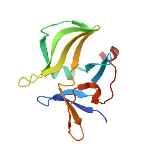
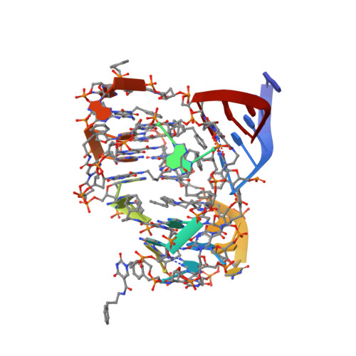
Evolving A RIG-I Antagonist: A Modified DNA Aptamer Mimics Viral RNA.

Ren, X.Gelinas, A.D.Linehan, M.Iwasaki, A.Wang, W.Janjic, N.Pyle, A.M.

(2021) J Mol Biology 433: 167227-167227

  • DOI: https://doi.org/10.1016/j.jmb.2021.167227
  • Primary Citation Related Structures: 
    7MK1

  • PubMed Abstract: 

    Vertebrate organisms express a diversity of protein receptors that recognize and respond to the presence of pathogenic molecules, functioning as an early warning system for infection. As a result of mutation or dysregulated metabolism, these same innate immune receptors can be inappropriately activated, leading to inflammation and disease. One of the most important receptors for detection and response to RNA viruses is called RIG-I, and dysregulation of this protein is linked with a variety of disease states. Despite its central role in inflammatory responses, antagonists for RIG-I are underdeveloped. In this study, we use invitro selection from a pool of modified DNA aptamers to create a high affinity RIG-I antagonist. A high resolution crystal structure of the complex reveals molecular mimicry between the aptamer and the 5'-triphosphate terminus of viral ligands, which bind to the same amino acids within the CTD recognition platform of the RIG-I receptor. Our study suggests a powerful, generalizable strategy for generating immunomodulatory drugs and mechanistic tool compounds.


  • Organizational Affiliation
    • Department of Molecular, Cellular and Developmental Biology, Yale University, New Haven, CT 06511, USA; Howard Hughes Medical Institute, Yale University, New Haven, CT 06520, USA.

Macromolecule Content 

  • Total Structure Weight: 59.58 kDa 
  • Atom Count: 4,454 
  • Modeled Residue Count: 324 
  • Deposited Residue Count: 332 
  • Unique protein chains: 1
  • Unique nucleic acid chains: 1

Macromolecules


Find similar proteins by:|  3D Structure
Entity ID: 1
MoleculeChains  Sequence LengthOrganismDetailsImage
Antiviral innate immune response receptor RIG-I
A, B
125Homo sapiensMutation(s): 0 
Gene Names: DDX58
EC: 3.6.4.13
UniProt & NIH Common Fund Data Resources
Find proteins for O95786 (Homo sapiens)
Explore O95786 
Go to UniProtKB:  O95786
PHAROS:  O95786
GTEx:  ENSG00000107201 
Entity Groups
Sequence Clusters30% Identity50% Identity70% Identity90% Identity95% Identity100% Identity
UniProt GroupO95786
Sequence Annotations
Expand
Reference Sequence
Find similar nucleic acids by:  (by identity cutoff) 
Entity ID: 2
MoleculeChains LengthOrganismImage
DNA (41-MER)
C, D
41synthetic construct
Sequence Annotations
Expand
Reference Sequence

Experimental Data & Validation

Experimental Data

  • Method: X-RAY DIFFRACTION
  • Resolution: 1.90 Å
  • R-Value Free:  0.220 (Depositor), 0.229 (DCC) 
  • R-Value Work:  0.188 (Depositor), 0.193 (DCC) 
  • R-Value Observed: 0.190 (Depositor) 
Space Group: P 21 21 21
Unit Cell:
Length ( Å )Angle ( ˚ )
a = 77.126α = 90
b = 86.632β = 90
c = 87.126γ = 90
Software Package:
Software NamePurpose
PHENIXrefinement
XDSdata reduction
Aimlessdata scaling
PHENIXphasing

Structure Validation

View Full Validation Report


Entry History 

& Funding Information

Deposition Data


Funding OrganizationLocationGrant Number
Howard Hughes Medical Institute (HHMI)United States--

Revision History  (Full details and data files)

  • Version 1.0: 2021-11-03
    Type: Initial release
  • Version 1.1: 2023-10-18
    Changes: Data collection, Refinement description