7MDU | pdb_00007mdu

BG505 SOSIP MD39 in complex with the monoclonal antibodies Rh.33104 mAb.1 and RM20A3


Experimental Data Snapshot

  • Method: ELECTRON MICROSCOPY
  • Resolution: 3.30 Å
  • Aggregation State: PARTICLE 
  • Reconstruction Method: SINGLE PARTICLE 

Starting Models: experimental
View more details

wwPDB Validation 3D Report Full Report

Validation slider image for 7MDU

This is version 1.2 of the entry. See complete history

Literature

From structure to sequence: Antibody discovery using cryoEM.

Antanasijevic, A.Bowman, C.A.Kirchdoerfer, R.N.Cottrell, C.A.Ozorowski, G.Upadhyay, A.A.Cirelli, K.M.Carnathan, D.G.Enemuo, C.A.Sewall, L.M.Nogal, B.Zhao, F.Groschel, B.Schief, W.R.Sok, D.Silvestri, G.Crotty, S.Bosinger, S.E.Ward, A.B.

(2022) Sci Adv 8: eabk2039-eabk2039

  • DOI: https://doi.org/10.1126/sciadv.abk2039
  • Primary Citation Related Structures: 
    7MDT, 7MDU, 7MEP

  • PubMed Abstract: 

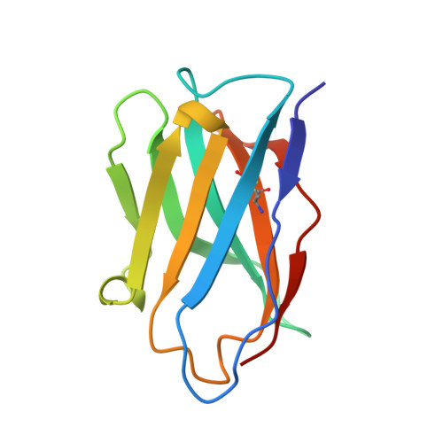
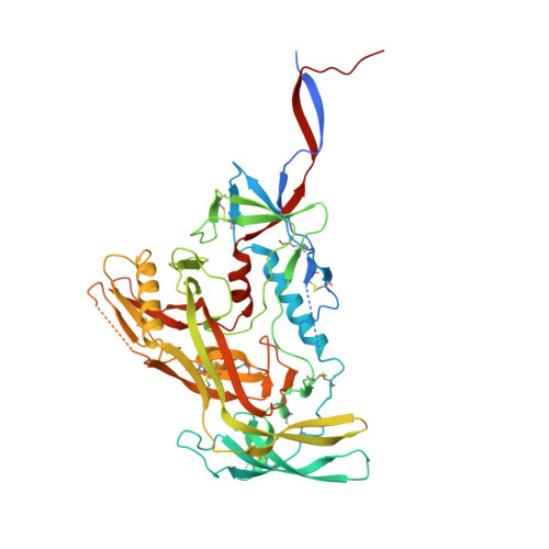
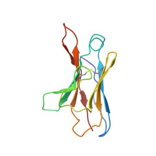
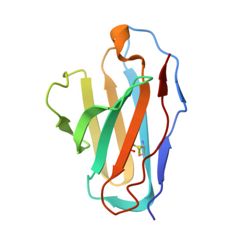
    One of the rate-limiting steps in analyzing immune responses to vaccines or infections is the isolation and characterization of monoclonal antibodies. Here, we present a hybrid structural and bioinformatic approach to directly assign the heavy and light chains, identify complementarity-determining regions, and discover sequences from cryoEM density maps of serum-derived polyclonal antibodies bound to an antigen. When combined with next-generation sequencing of immune repertoires, we were able to specifically identify clonal family members, synthesize the monoclonal antibodies, and confirm that they interact with the antigen in a manner equivalent to the corresponding polyclonal antibodies. This structure-based approach for identification of monoclonal antibodies from polyclonal sera opens new avenues for analysis of immune responses and iterative vaccine design.


  • Organizational Affiliation
    • Department of Integrative Structural and Computational Biology, The Scripps Research Institute, La Jolla, CA 92037, USA.

Macromolecule Content 

  • Total Structure Weight: 130.96 kDa 
  • Atom Count: 8,332 
  • Modeled Residue Count: 1,018 
  • Deposited Residue Count: 1,124 
  • Unique protein chains: 6

Macromolecules

Find similar proteins by:|  3D Structure
Entity ID: 1
MoleculeChains  Sequence LengthOrganismDetailsImage
Rh.33104 mAb.1 Heavy ChainA [auth H]116Macaca mulattaMutation(s): 0 
Find similar proteins by:|  3D Structure
Entity ID: 2
MoleculeChains  Sequence LengthOrganismDetailsImage
Surface protein gp120B [auth A]513Human immunodeficiency virus 1Mutation(s): 0 
Gene Names: env
UniProt
Find proteins for Q2N0S6 (Human immunodeficiency virus type 1)
Explore Q2N0S6 
Go to UniProtKB:  Q2N0S6
Entity Groups
Sequence Clusters30% Identity50% Identity70% Identity90% Identity95% Identity100% Identity
UniProt GroupQ2N0S6
Glycosylation
Glycosylation Sites: 18
Sequence Annotations
Expand
Reference Sequence
Find similar proteins by:|  3D Structure
Entity ID: 3
MoleculeChains  Sequence LengthOrganismDetailsImage
RM20A3 mAb Heavy ChainC [auth E]125Macaca mulattaMutation(s): 0 
Find similar proteins by:|  3D Structure
Entity ID: 4
MoleculeChains  Sequence LengthOrganismDetailsImage
RM20A3 mAb Light ChainD [auth F]111Macaca mulattaMutation(s): 0 
Find similar proteins by:|  3D Structure
Entity ID: 5
MoleculeChains  Sequence LengthOrganismDetailsImage
Rh.33104 mAb.1 Light ChainE [auth L]106Macaca mulattaMutation(s): 0 
Find similar proteins by:|  3D Structure
Entity ID: 6
MoleculeChains  Sequence LengthOrganismDetailsImage
Transmembrane protein gp41F [auth B]153Human immunodeficiency virus 1Mutation(s): 0 
Gene Names: env
UniProt
Find proteins for Q2N0S6 (Human immunodeficiency virus type 1)
Explore Q2N0S6 
Go to UniProtKB:  Q2N0S6
Entity Groups
Sequence Clusters30% Identity50% Identity70% Identity90% Identity95% Identity100% Identity
UniProt GroupQ2N0S6
Glycosylation
Glycosylation Sites: 3
Sequence Annotations
Expand
Reference Sequence

Oligosaccharides

Help  
Entity ID: 7
MoleculeChains Length2D Diagram GlycosylationD Interactions
2-acetamido-2-deoxy-beta-D-glucopyranose-(1-4)-2-acetamido-2-deoxy-beta-D-glucopyranose
G [auth C],
H [auth D],
I [auth G],
K [auth J],
L [auth K],
G [auth C],
H [auth D],
I [auth G],
K [auth J],
L [auth K],
M,
N,
O
2N-Glycosylation
Glycosylation Resources
GlyTouCan: G42666HT
GlyCosmos: G42666HT
GlyGen: G42666HT
Entity ID: 8
MoleculeChains Length2D Diagram GlycosylationD Interactions
alpha-D-mannopyranose-(1-3)-[alpha-D-mannopyranose-(1-6)]beta-D-mannopyranose-(1-4)-2-acetamido-2-deoxy-beta-D-glucopyranose-(1-4)-2-acetamido-2-deoxy-beta-D-glucopyranoseJ [auth I]5N-Glycosylation
Glycosylation Resources
GlyTouCan: G22768VO
GlyCosmos: G22768VO
GlyGen: G22768VO

Small Molecules

Ligands 1 Unique
IDChains Name / Formula / InChI Key2D Diagram3D Interactions
NAG

Query on NAG


Download:Ideal Coordinates CCD File
AA [auth B]
P [auth A]
Q [auth A]
R [auth A]
S [auth A]
AA [auth B],
P [auth A],
Q [auth A],
R [auth A],
S [auth A],
T [auth A],
U [auth A],
V [auth A],
W [auth A],
X [auth A],
Y [auth B],
Z [auth B]
2-acetamido-2-deoxy-beta-D-glucopyranose
C8 H15 N O6
OVRNDRQMDRJTHS-FMDGEEDCSA-N

Experimental Data & Validation

Experimental Data

  • Method: ELECTRON MICROSCOPY
  • Resolution: 3.30 Å
  • Aggregation State: PARTICLE 
  • Reconstruction Method: SINGLE PARTICLE 
EM Software:
TaskSoftware PackageVersion
RECONSTRUCTIONRELION3.0
MODEL REFINEMENTRosettaEM

Structure Validation

View Full Validation Report


Entry History 

& Funding Information

Deposition Data


Funding OrganizationLocationGrant Number
Bill & Melinda Gates FoundationUnited StatesOPP1115782
Bill & Melinda Gates FoundationUnited StatesOPP1196345

Revision History  (Full details and data files)

  • Version 1.0: 2022-01-26
    Type: Initial release
  • Version 1.1: 2022-02-02
    Changes: Database references
  • Version 1.2: 2024-11-06
    Changes: Data collection, Refinement description, Structure summary