7MCS | pdb_00007mcs

Cryo-electron microscopy structure of TnsC(1-503)A225V bound to DNA


Experimental Data Snapshot

  • Method: ELECTRON MICROSCOPY
  • Resolution: 3.56 Å
  • Aggregation State: PARTICLE 
  • Reconstruction Method: SINGLE PARTICLE 

wwPDB Validation 3D Report Full Report

Validation slider image for 7MCS

This is version 1.2 of the entry. See complete history

Literature

Structural basis for DNA targeting by the Tn7 transposon.

Shen, Y.Gomez-Blanco, J.Petassi, M.T.Peters, J.E.Ortega, J.Guarne, A.

(2022) Nat Struct Mol Biol 29: 143-151

  • DOI: https://doi.org/10.1038/s41594-022-00724-8
  • Primary Citation Related Structures: 
    7MBW, 7MCS

  • PubMed Abstract: 

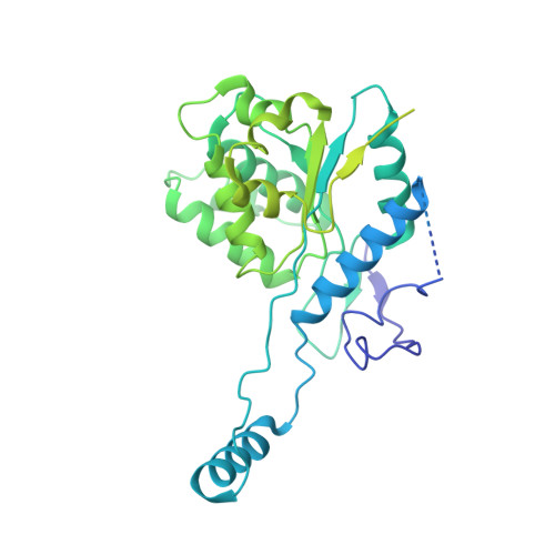
    Tn7 transposable elements are unique for their highly specific, and sometimes programmable, target-site selection mechanisms and precise insertions. All the elements in the Tn7 family utilize an AAA+ adaptor (TnsC) to coordinate target-site selection with transpososome assembly and to prevent insertions at sites already containing a Tn7 element. Owing to its multiple functions, TnsC is considered the linchpin in the Tn7 element. Here we present the high-resolution cryo-EM structure of TnsC bound to DNA using a gain-of-function variant of the protein and a DNA substrate that together recapitulate the recruitment to a specific DNA target site. TnsC forms an asymmetric ring on target DNA that segregates target-site selection and interaction with the paired-end complex to opposite faces of the ring. Unlike most AAA+ ATPases, TnsC uses a DNA distortion to find the target site but does not remodel DNA to activate transposition. By recognizing pre-distorted substrates, TnsC creates a built-in regulatory mechanism where ATP hydrolysis abolishes ring formation proximal to an existing element. This work unveils how Tn7 and Tn7-like elements determine the strict spacing between the target and integration sites.


  • Organizational Affiliation
    • Department of Biochemistry, McGill University, Montreal, Quebec, Canada.

Macromolecule Content 

  • Total Structure Weight: 427.8 kDa 
  • Atom Count: 21,600 
  • Modeled Residue Count: 2,649 
  • Deposited Residue Count: 3,691 
  • Unique protein chains: 1
  • Unique nucleic acid chains: 2

Macromolecules


Find similar proteins by:|  3D Structure
Entity ID: 1
MoleculeChains  Sequence LengthOrganismDetailsImage
Transposon Tn7 transposition protein TnsC
A, B, C, D, E
A, B, C, D, E, F, G
523Escherichia coliMutation(s): 2 
Gene Names: tnsC
UniProt
Find proteins for P05846 (Escherichia coli)
Explore P05846 
Go to UniProtKB:  P05846
Entity Groups
Sequence Clusters30% Identity50% Identity70% Identity90% Identity95% Identity100% Identity
UniProt GroupP05846
Sequence Annotations
Expand
Reference Sequence
Find similar nucleic acids by:  Sequence
Entity ID: 2
MoleculeChains LengthOrganismImage
DNA (5'-D(P*CP*GP*CP*GP*CP*GP*CP*GP*CP*GP*CP*GP*CP*GP*C)-3')15synthetic construct
Sequence Annotations
Expand
Reference Sequence
Find similar nucleic acids by:  Sequence
Entity ID: 3
MoleculeChains LengthOrganismImage
DNA (5'-D(P*GP*CP*GP*CP*GP*CP*GP*CP*GP*CP*GP*CP*GP*CP*G)-3')15synthetic construct
Sequence Annotations
Expand
Reference Sequence

Small Molecules

Ligands 2 Unique
IDChains Name / Formula / InChI Key2D Diagram3D Interactions
ANP
(Subject of Investigation/LOI)

Query on ANP


Download:Ideal Coordinates CCD File
J [auth B]
L [auth C]
N [auth D]
P [auth E]
R [auth F]
J [auth B],
L [auth C],
N [auth D],
P [auth E],
R [auth F],
T [auth G]
PHOSPHOAMINOPHOSPHONIC ACID-ADENYLATE ESTER
C10 H17 N6 O12 P3
PVKSNHVPLWYQGJ-KQYNXXCUSA-N
MG
(Subject of Investigation/LOI)

Query on MG


Download:Ideal Coordinates CCD File
K [auth B]
M [auth C]
O [auth D]
Q [auth E]
S [auth F]
K [auth B],
M [auth C],
O [auth D],
Q [auth E],
S [auth F],
U [auth G]
MAGNESIUM ION
Mg
JLVVSXFLKOJNIY-UHFFFAOYSA-N

Experimental Data & Validation

Experimental Data

  • Method: ELECTRON MICROSCOPY
  • Resolution: 3.56 Å
  • Aggregation State: PARTICLE 
  • Reconstruction Method: SINGLE PARTICLE 
EM Software:
TaskSoftware PackageVersion
RECONSTRUCTIONRELION3.1
MODEL REFINEMENTPHENIX

Structure Validation

View Full Validation Report


Entry History 

& Funding Information

Deposition Data


Funding OrganizationLocationGrant Number
Canadian Institutes of Health Research (CIHR)CanadaPJT-155941

Revision History  (Full details and data files)

  • Version 1.0: 2022-02-23
    Type: Initial release
  • Version 1.1: 2022-03-02
    Changes: Database references
  • Version 1.2: 2024-05-29
    Changes: Data collection