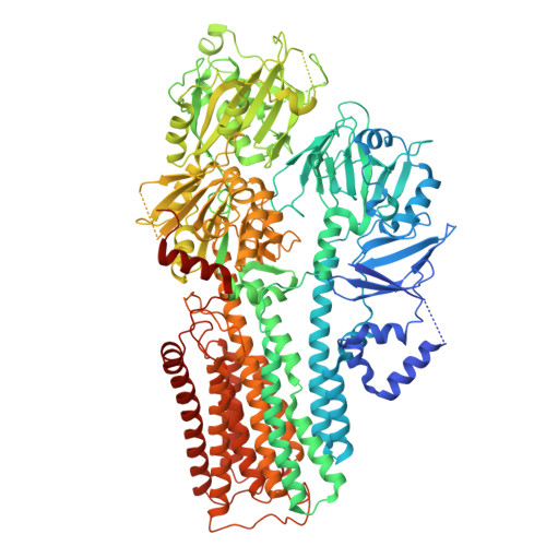
7M5V

human ATP13A2 in the AMPPNP-bound occluded state


Experimental Data Snapshot

  • Method: ELECTRON MICROSCOPY
  • Resolution: 2.90 Å
  • Aggregation State: PARTICLE 
  • Reconstruction Method: SINGLE PARTICLE 

wwPDB Validation   3D Report Full Report


This is version 2.0 of the entry. See complete history


Literature

Structural mechanisms for gating and ion selectivity of the human polyamine transporter ATP13A2.

Tillinghast, J.Drury, S.Bowser, D.Benn, A.Lee, K.P.K.

(2021) Mol Cell 81: 4650-4662.e4

  • DOI: https://doi.org/10.1016/j.molcel.2021.10.002
  • Primary Citation of Related Structures:  
    7M5V, 7M5X, 7M5Y

  • PubMed Abstract: 

    Mutations in ATP13A2, also known as PARK9, cause a rare monogenic form of juvenile-onset Parkinson's disease named Kufor-Rakeb syndrome and other neurodegenerative diseases. ATP13A2 encodes a neuroprotective P5B P-type ATPase highly enriched in the brain that mediates selective import of spermine ions from lysosomes into the cytosol via an unknown mechanism. Here we present three structures of human ATP13A2 bound to an ATP analog or to spermine in the presence of phosphomimetics determined by cryoelectron microscopy. ATP13A2 autophosphorylation opens a lysosome luminal gate to reveal a narrow lumen access channel that holds a spermine ion in its entrance. ATP13A2's architecture suggests physical principles underlying selective polyamine transport and anticipates a "pump-channel" intermediate that could function as a counter-cation conduit to facilitate lysosome acidification. Our findings establish a firm foundation to understand ATP13A2 mutations associated with disease and bring us closer to realizing ATP13A2's potential in neuroprotective therapy.


  • Organizational Affiliation

    Department of Cellular and Molecular Physiology, Pennsylvania State University College of Medicine, The Pennsylvania State University, Hershey, PA 17033, USA.


Macromolecules
Find similar proteins by:  (by identity cutoff)  |  3D Structure
Entity ID: 1
MoleculeChains Sequence LengthOrganismDetailsImage
Polyamine-transporting ATPase 13A21,188Homo sapiensMutation(s): 0 
Gene Names: ATP13A2PARK9
EC: 7.6.2
Membrane Entity: Yes 
UniProt & NIH Common Fund Data Resources
Find proteins for Q9NQ11 (Homo sapiens)
Explore Q9NQ11 
Go to UniProtKB:  Q9NQ11
PHAROS:  Q9NQ11
GTEx:  ENSG00000159363 
Entity Groups  
Sequence Clusters30% Identity50% Identity70% Identity90% Identity95% Identity100% Identity
UniProt GroupQ9NQ11
Glycosylation
Glycosylation Sites: 1Go to GlyGen: Q9NQ11-1
Sequence Annotations
Expand
  • Reference Sequence
Small Molecules
Ligands 9 Unique
IDChains Name / Formula / InChI Key2D Diagram3D Interactions
EUJ (Subject of Investigation/LOI)
Query on EUJ

Download Ideal Coordinates CCD File 
Y [auth A](2R)-3-{[(S)-hydroxy{[(1S,2R,3R,4S,5S,6R)-2,4,6-trihydroxy-3,5-bis(phosphonooxy)cyclohexyl]oxy}phosphoryl]oxy}propane-1,2-diyl dioctanoate
C25 H49 O19 P3
QXHVLVSULWMTCV-DICZBTHZSA-N
LMT
Query on LMT

Download Ideal Coordinates CCD File 
D [auth A]DODECYL-BETA-D-MALTOSIDE
C24 H46 O11
NLEBIOOXCVAHBD-QKMCSOCLSA-N
ANP (Subject of Investigation/LOI)
Query on ANP

Download Ideal Coordinates CCD File 
X [auth A]PHOSPHOAMINOPHOSPHONIC ACID-ADENYLATE ESTER
C10 H17 N6 O12 P3
PVKSNHVPLWYQGJ-KQYNXXCUSA-N
Y01
Query on Y01

Download Ideal Coordinates CCD File 
E [auth A],
F [auth A],
K [auth A],
L [auth A]
CHOLESTEROL HEMISUCCINATE
C31 H50 O4
WLNARFZDISHUGS-MIXBDBMTSA-N
NAG
Query on NAG

Download Ideal Coordinates CCD File 
S [auth A]2-acetamido-2-deoxy-beta-D-glucopyranose
C8 H15 N O6
OVRNDRQMDRJTHS-FMDGEEDCSA-N
1DO
Query on 1DO

Download Ideal Coordinates CCD File 
C [auth A],
I [auth A],
J [auth A],
R [auth A]
1-DODECANOL
C12 H26 O
LQZZUXJYWNFBMV-UHFFFAOYSA-N
D12
Query on D12

Download Ideal Coordinates CCD File 
H [auth A],
M [auth A]
DODECANE
C12 H26
SNRUBQQJIBEYMU-UHFFFAOYSA-N
D10
Query on D10

Download Ideal Coordinates CCD File 
G [auth A]
N [auth A]
O [auth A]
P [auth A]
Q [auth A]
G [auth A],
N [auth A],
O [auth A],
P [auth A],
Q [auth A],
T [auth A],
U [auth A],
V [auth A],
W [auth A]
DECANE
C10 H22
DIOQZVSQGTUSAI-UHFFFAOYSA-N
MG (Subject of Investigation/LOI)
Query on MG

Download Ideal Coordinates CCD File 
B [auth A]MAGNESIUM ION
Mg
JLVVSXFLKOJNIY-UHFFFAOYSA-N
Experimental Data & Validation

Experimental Data

  • Method: ELECTRON MICROSCOPY
  • Resolution: 2.90 Å
  • Aggregation State: PARTICLE 
  • Reconstruction Method: SINGLE PARTICLE 

Structure Validation

View Full Validation Report


Entry History & Funding Information

Deposition Data

  • Released Date: 2022-03-30 
  • Deposition Author(s): Lee, K.P.K.

Funding OrganizationLocationGrant Number
Not funded--

Revision History  (Full details and data files)

  • Version 1.0: 2022-03-30
    Type: Initial release
  • Version 2.0: 2022-04-27
    Type: Coordinate replacement
    Reason: Chirality error
    Changes: Advisory, Atomic model, Author supporting evidence, Data collection, Database references, Derived calculations, Polymer sequence, Source and taxonomy, Structure summary