7M3Y | pdb_00007m3y

Structure of TIM-3 in complex with 8-chloro-2-methyl-9-(3-mehtylpyridin-4-yl)-[1,2,4]triazolo[1,5-c]quinazolin-5(6H)-one (compound 22)


Experimental Data Snapshot

  • Method: X-RAY DIFFRACTION
  • Resolution: 1.69 Å
  • R-Value Free: 
    0.213 (Depositor), 0.211 (DCC) 
  • R-Value Work: 
    0.187 (Depositor), 0.186 (DCC) 
  • R-Value Observed: 
    0.188 (Depositor) 

Starting Model: experimental
View more details

wwPDB Validation 3D Report Full Report

Validation slider image for 7M3Y

Ligand Structure Quality Assessment 


This is version 1.3 of the entry. See complete history

Literature

Fragment-Based Discovery of Small Molecules Bound to T-Cell Immunoglobulin and Mucin Domain-Containing Molecule 3 (TIM-3).

Rietz, T.A.Teuscher, K.B.Mills, J.J.Gogliotti, R.D.Lepovitz, L.T.Scaggs, W.R.Yoshida, K.Luong, K.Lee, T.Fesik, S.W.

(2021) J Med Chem 64: 14757-14772

  • DOI: https://doi.org/10.1021/acs.jmedchem.1c01336
  • Primary Citation Related Structures: 
    7M3Y, 7M3Z, 7M41

  • PubMed Abstract: 

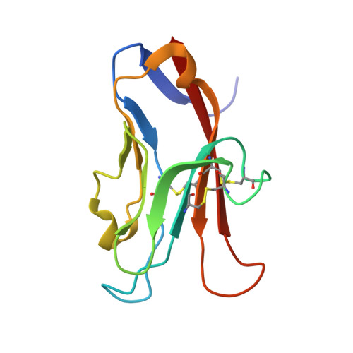
    T-cell immunoglobulin and mucin domain-containing molecule 3 (TIM-3; HAVCR2) has emerged as an attractive immune checkpoint target for cancer immunotherapy. TIM-3 is a negative regulator of the systemic immune response to cancer and is expressed on several dysfunctional, or exhausted, immune cell subsets. Upregulation of TIM-3 is associated with tumor progression, poor survival rates, and acquired resistance to antibody-based immunotherapies in the clinic. Despite the potential advantages of small-molecule inhibitors over antibodies, the discovery of small-molecule inhibitors has lagged behind that of antibody therapeutics. Here, we describe the discovery of high-affinity small-molecule ligands for TIM-3 through an NMR-based fragment screen and structure-based lead optimization. These compounds represent useful tools to further study the biology of TIM-3 immune modulation in cancer and serve as a potentially useful starting point toward the discovery of TIM-3-targeted therapeutics.


  • Organizational Affiliation
    • Department of Biochemistry, Vanderbilt University School of Medicine, Nashville, Tennessee 37232-0146, United States.

Macromolecule Content 

  • Total Structure Weight: 12.66 kDa 
  • Atom Count: 997 
  • Modeled Residue Count: 109 
  • Deposited Residue Count: 109 
  • Unique protein chains: 1

Macromolecules

Find similar proteins by:|  3D Structure
Entity ID: 1
MoleculeChains  Sequence LengthOrganismDetailsImage
Hepatitis A virus cellular receptor 2A [auth B]109Homo sapiensMutation(s): 0 
Gene Names: HAVCR2TIM3TIMD3
Membrane Entity: Yes 
UniProt & NIH Common Fund Data Resources
Find proteins for Q8TDQ0 (Homo sapiens)
Explore Q8TDQ0 
Go to UniProtKB:  Q8TDQ0
PHAROS:  Q8TDQ0
GTEx:  ENSG00000135077 
Entity Groups
Sequence Clusters30% Identity50% Identity70% Identity90% Identity95% Identity100% Identity
UniProt GroupQ8TDQ0
Sequence Annotations
Expand
Reference Sequence

Small Molecules

Ligands 2 Unique
IDChains Name / Formula / InChI Key2D Diagram3D Interactions
YQ7
(Subject of Investigation/LOI)

Query on YQ7


Download:Ideal Coordinates CCD File
B
(4R,10aP)-8-chloro-2-methyl-9-(3-methylpyridin-4-yl)[1,2,4]triazolo[1,5-c]quinazolin-5(6H)-one
C16 H12 Cl N5 O
FVTXWRGKBOWNPA-UHFFFAOYSA-N
CA

Query on CA


Download:Ideal Coordinates CCD File
C [auth B]CALCIUM ION
Ca
BHPQYMZQTOCNFJ-UHFFFAOYSA-N

Experimental Data & Validation

Experimental Data

  • Method: X-RAY DIFFRACTION
  • Resolution: 1.69 Å
  • R-Value Free:  0.213 (Depositor), 0.211 (DCC) 
  • R-Value Work:  0.187 (Depositor), 0.186 (DCC) 
  • R-Value Observed: 0.188 (Depositor) 
Space Group: C 2 2 21
Unit Cell:
Length ( Å )Angle ( ˚ )
a = 46.958α = 90
b = 85.343β = 90
c = 53.697γ = 90
Software Package:
Software NamePurpose
HKL-2000data scaling
PHASERphasing
PHENIXrefinement
PDB_EXTRACTdata extraction
HKL-2000data reduction

Structure Validation

View Full Validation Report


Ligand Structure Quality Assessment 


Entry History 

Deposition Data

  • Released Date: 2021-10-13 
  • Deposition Author(s): Rietz, T.A.

Revision History  (Full details and data files)

  • Version 1.0: 2021-10-13
    Type: Initial release
  • Version 1.1: 2021-10-27
    Changes: Database references
  • Version 1.2: 2023-10-18
    Changes: Data collection, Refinement description
  • Version 1.3: 2024-11-13
    Changes: Structure summary