7M3F

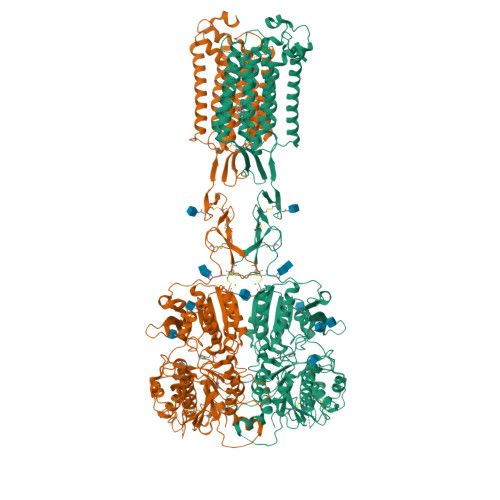
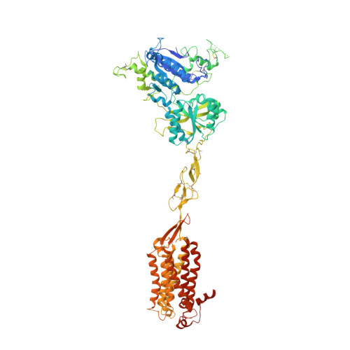
Asymmetric Activation of the Calcium Sensing Receptor Homodimer


Experimental Data Snapshot

  • Method: ELECTRON MICROSCOPY
  • Resolution: 2.80 Å
  • Aggregation State: PARTICLE 
  • Reconstruction Method: SINGLE PARTICLE 

wwPDB Validation   3D Report Full Report


This is version 1.3 of the entry. See complete history


Literature

Asymmetric activation of the calcium-sensing receptor homodimer.

Gao, Y.Robertson, M.J.Rahman, S.N.Seven, A.B.Zhang, C.Meyerowitz, J.G.Panova, O.Hannan, F.M.Thakker, R.V.Brauner-Osborne, H.Mathiesen, J.M.Skiniotis, G.

(2021) Nature 595: 455-459

  • DOI: https://doi.org/10.1038/s41586-021-03691-0
  • Primary Citation of Related Structures:  
    7M3E, 7M3F, 7M3G, 7M3J

  • PubMed Abstract: 

    The calcium-sensing receptor (CaSR), a cell-surface sensor for Ca 2+ , is the master regulator of calcium homeostasis in humans and is the target of calcimimetic drugs for the treatment of parathyroid disorders 1 . CaSR is a family C G-protein-coupled receptor 2 that functions as an obligate homodimer, with each protomer composed of a Ca 2+ -binding extracellular domain and a seven-transmembrane-helix domain (7TM) that activates heterotrimeric G proteins. Here we present cryo-electron microscopy structures of near-full-length human CaSR in inactive or active states bound to Ca 2+ and various calcilytic or calcimimetic drug molecules. We show that, upon activation, the CaSR homodimer adopts an asymmetric 7TM configuration that primes one protomer for G-protein coupling. This asymmetry is stabilized by 7TM-targeting calcimimetic drugs adopting distinctly different poses in the two protomers, whereas the binding of a calcilytic drug locks CaSR 7TMs in an inactive symmetric configuration. These results provide a detailed structural framework for CaSR activation and the rational design of therapeutics targeting this receptor.


  • Organizational Affiliation

    Department of Molecular and Cellular Physiology, Stanford University School of Medicine, Stanford, CA, USA.


Macromolecules
Find similar proteins by:  (by identity cutoff)  |  3D Structure
Entity ID: 1
MoleculeChains Sequence LengthOrganismDetailsImage
Extracellular calcium-sensing receptor
A, B
902Homo sapiensMutation(s): 0 
Gene Names: CASRGPRC2APCAR1
Membrane Entity: Yes 
UniProt & NIH Common Fund Data Resources
Find proteins for P41180 (Homo sapiens)
Explore P41180 
Go to UniProtKB:  P41180
PHAROS:  P41180
GTEx:  ENSG00000036828 
Entity Groups  
Sequence Clusters30% Identity50% Identity70% Identity90% Identity95% Identity100% Identity
UniProt GroupP41180
Glycosylation
Glycosylation Sites: 6Go to GlyGen: P41180-1
Sequence Annotations
Expand
  • Reference Sequence
Oligosaccharides

Help

Entity ID: 2
MoleculeChains Length2D Diagram Glycosylation3D Interactions
2-acetamido-2-deoxy-beta-D-glucopyranose-(1-4)-2-acetamido-2-deoxy-beta-D-glucopyranose
C, D, E
2N-Glycosylation
Glycosylation Resources
GlyTouCan:  G42666HT
GlyCosmos:  G42666HT
GlyGen:  G42666HT
Small Molecules
Ligands 5 Unique
IDChains Name / Formula / InChI Key2D Diagram3D Interactions
YP4 (Subject of Investigation/LOI)
Query on YP4

Download Ideal Coordinates CCD File 
J [auth A],
U [auth B]
N-[(1R)-1-(naphthalen-1-yl)ethyl]-3-[3-(trifluoromethyl)phenyl]propan-1-amine
C22 H22 F3 N
VDHAWDNDOKGFTD-MRXNPFEDSA-N
NAG
Query on NAG

Download Ideal Coordinates CCD File 
F [auth A]
G [auth A]
H [auth A]
I [auth A]
P [auth B]
F [auth A],
G [auth A],
H [auth A],
I [auth A],
P [auth B],
Q [auth B],
R [auth B],
S [auth B],
T [auth B]
2-acetamido-2-deoxy-beta-D-glucopyranose
C8 H15 N O6
OVRNDRQMDRJTHS-FMDGEEDCSA-N
TRP
Query on TRP

Download Ideal Coordinates CCD File 
K [auth A],
V [auth B]
TRYPTOPHAN
C11 H12 N2 O2
QIVBCDIJIAJPQS-VIFPVBQESA-N
PO4
Query on PO4

Download Ideal Coordinates CCD File 
O [auth A],
X [auth B]
PHOSPHATE ION
O4 P
NBIIXXVUZAFLBC-UHFFFAOYSA-K
CA
Query on CA

Download Ideal Coordinates CCD File 
L [auth A],
M [auth A],
N [auth A],
W [auth B]
CALCIUM ION
Ca
BHPQYMZQTOCNFJ-UHFFFAOYSA-N
Binding Affinity Annotations 
IDSourceBinding Affinity
YP4 BindingDB:  7M3F EC50: min: 19.95, max: 180 (nM) from 3 assay(s)
Experimental Data & Validation

Experimental Data

  • Method: ELECTRON MICROSCOPY
  • Resolution: 2.80 Å
  • Aggregation State: PARTICLE 
  • Reconstruction Method: SINGLE PARTICLE 
EM Software:
TaskSoftware PackageVersion
RECONSTRUCTIONSerialEM

Structure Validation

View Full Validation Report


Entry History & Funding Information

Deposition Data


Funding OrganizationLocationGrant Number
National Institutes of Health/National Institute of Neurological Disorders and Stroke (NIH/NINDS)United StatesR01NS092695

Revision History  (Full details and data files)

  • Version 1.0: 2021-06-30
    Type: Initial release
  • Version 1.1: 2021-07-14
    Changes: Database references
  • Version 1.2: 2021-07-21
    Changes: Database references
  • Version 1.3: 2024-11-20
    Changes: Data collection, Database references, Structure summary