Chemoproteomic identification of CO 2 -dependent lysine carboxylation in proteins.
King, D.T., Zhu, S., Hardie, D.B., Serrano-Negron, J.E., Madden, Z., Kolappan, S., Vocadlo, D.J.(2022) Nat Chem Biol 18: 782-791
- PubMed: 35710617 Search on PubMedSearch on PubMed Central
- DOI: https://doi.org/10.1038/s41589-022-01043-1
- Primary Citation Related Structures:
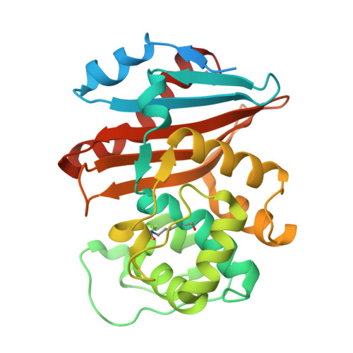
7LXG - PubMed Abstract:
Carbon dioxide is an omnipresent gas that drives adaptive responses within organisms from all domains of life. The molecular mechanisms by which proteins serve as sensors of CO 2 are, accordingly, of great interest. Because CO 2 is electrophilic, one way it can modulate protein biochemistry is by carboxylation of the amine group of lysine residues. However, the resulting CO 2 -carboxylated lysines spontaneously decompose, giving off CO 2 , which makes studying this modification difficult. Here we describe a method to stably mimic CO 2 -carboxylated lysine residues in proteins. We leverage this method to develop a quantitative approach to identify CO 2 -carboxylated lysines of proteins and explore the lysine 'carboxylome' of the CO 2 -responsive cyanobacterium Synechocystis sp. We uncover one CO 2 -carboxylated lysine within the effector binding pocket of the metabolic signaling protein PII. CO 2 -carboxylatation of this lysine markedly lowers the affinity of PII for its regulatory effector ligand ATP, illuminating a negative molecular control mechanism mediated by CO 2 .
- Department of Molecular Biology and Biochemistry, Simon Fraser University, Burnaby, British Columbia, Canada.
Organizational Affiliation: