7LWB

Crystal Structure of phospho-Rab8a with the RH2 domain (117-165) of RILPL2


Experimental Data Snapshot

  • Method: X-RAY DIFFRACTION
  • Resolution: 1.90 Å
  • R-Value Free: 0.267 
  • R-Value Work: 0.219 
  • R-Value Observed: 0.221 

wwPDB Validation   3D Report Full Report


Ligand Structure Quality Assessment 


This is version 1.3 of the entry. See complete history


Literature

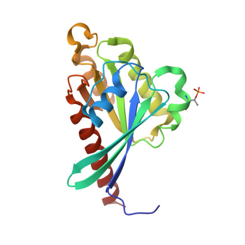
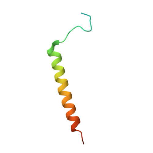
Dual arginine recognition of LRRK2 phosphorylated Rab GTPases.

Waschbusch, D.Purlyte, E.Khan, A.R.

(2021) Biophys J 120: 1846-1855

  • DOI: https://doi.org/10.1016/j.bpj.2021.03.030
  • Primary Citation of Related Structures:  
    6WHE, 7LWB

  • PubMed Abstract: 

    Parkinson's-disease-associated LRRK2 is a multidomain Ser/Thr kinase that phosphorylates a subset of Rab GTPases to control their effector functions. Rab GTPases are the prime regulators of membrane trafficking in eukaryotic cells. Rabs exert their biological effects by recruitment of effector proteins to subcellular compartments via their Rab-binding domain (RBD). Effectors are modular and typically contain additional domains that regulate various aspects of vesicle formation, trafficking, fusion, and organelle dynamics. The RBD of effectors is typically an α-helical coiled coil that recognizes the GTP conformation of the switch 1 and switch 2 motifs of Rabs. LRRK2 phosphorylates Rab8a at T72 (pT72) of its switch 2 α-helix. This post-translational modification enables recruitment of RILPL2, an effector that regulates ciliogenesis in model cell lines. A newly identified RBD motif of RILPL2, termed the X-cap, has been shown to recognize the phosphate via direct interactions between an arginine residue (R132) and pT72 of Rab8a. Here, we show that a second distal arginine (R130) is also essential for phospho-Rab binding by RILPL2. Through structural, biophysical, and cellular studies, we find that R130 stabilizes the primary R132:pT72 salt bridge through favorable enthalpic contributions to the binding affinity. These findings may have implications for the mechanism by which LRRK2 activation leads to assembly of phospho-Rab complexes and subsequent control of their membrane trafficking functions in cells.


  • Organizational Affiliation

    School of Biochemistry and Immunology, Trinity College Dublin, Dublin, Ireland.


Macromolecules
Find similar proteins by:  (by identity cutoff)  |  3D Structure
Entity ID: 1
MoleculeChains Sequence LengthOrganismDetailsImage
Ras-related protein Rab-8A184Homo sapiensMutation(s): 1 
Gene Names: RAB8AMELRAB8
UniProt & NIH Common Fund Data Resources
Find proteins for P61006 (Homo sapiens)
Explore P61006 
Go to UniProtKB:  P61006
PHAROS:  P61006
GTEx:  ENSG00000167461 
Entity Groups  
Sequence Clusters30% Identity50% Identity70% Identity90% Identity95% Identity100% Identity
UniProt GroupP61006
Sequence Annotations
Expand
  • Reference Sequence
Find similar proteins by:  (by identity cutoff)  |  3D Structure
Entity ID: 2
MoleculeChains Sequence LengthOrganismDetailsImage
RILP-like protein 2B [auth D]53Homo sapiensMutation(s): 0 
Gene Names: RILPL2RLP2
UniProt & NIH Common Fund Data Resources
Find proteins for Q969X0 (Homo sapiens)
Explore Q969X0 
Go to UniProtKB:  Q969X0
PHAROS:  Q969X0
GTEx:  ENSG00000150977 
Entity Groups  
Sequence Clusters30% Identity50% Identity70% Identity90% Identity95% Identity100% Identity
UniProt GroupQ969X0
Sequence Annotations
Expand
  • Reference Sequence
Small Molecules
Ligands 2 Unique
IDChains Name / Formula / InChI Key2D Diagram3D Interactions
GTP
Query on GTP

Download Ideal Coordinates CCD File 
C [auth A]GUANOSINE-5'-TRIPHOSPHATE
C10 H16 N5 O14 P3
XKMLYUALXHKNFT-UUOKFMHZSA-N
MG
Query on MG

Download Ideal Coordinates CCD File 
D [auth A]MAGNESIUM ION
Mg
JLVVSXFLKOJNIY-UHFFFAOYSA-N
Modified Residues  1 Unique
IDChains TypeFormula2D DiagramParent
TPO
Query on TPO
A
L-PEPTIDE LINKINGC4 H10 N O6 PTHR
Experimental Data & Validation

Experimental Data

  • Method: X-RAY DIFFRACTION
  • Resolution: 1.90 Å
  • R-Value Free: 0.267 
  • R-Value Work: 0.219 
  • R-Value Observed: 0.221 
  • Space Group: C 2 2 21
Unit Cell:
Length ( Å )Angle ( ˚ )
a = 62.773α = 90
b = 68.43β = 90
c = 128.1γ = 90
Software Package:
Software NamePurpose
PHENIXrefinement
PDB_EXTRACTdata extraction
XDSdata reduction
Aimlessdata scaling
PHASERphasing

Structure Validation

View Full Validation Report


Ligand Structure Quality Assessment 


Entry History 

Deposition Data

Revision History  (Full details and data files)

  • Version 1.0: 2021-03-10
    Type: Initial release
  • Version 1.1: 2021-05-05
    Changes: Database references
  • Version 1.2: 2021-05-12
    Changes: Database references
  • Version 1.3: 2023-10-18
    Changes: Data collection, Database references, Refinement description