7LV3 | pdb_00007lv3

Crystal structure of human protein kinase G (PKG) R-C complex in inhibited state


Experimental Data Snapshot

  • Method: X-RAY DIFFRACTION
  • Resolution: 2.41 Å
  • R-Value Free: 
    0.238 (Depositor), 0.243 (DCC) 
  • R-Value Work: 
    0.200 (Depositor), 0.205 (DCC) 
  • R-Value Observed: 
    0.201 (Depositor) 

Starting Models: experimental
View more details

wwPDB Validation 3D Report Full Report

Validation slider image for 7LV3

Ligand Structure Quality Assessment 


This is version 1.2 of the entry. See complete history

Literature

An auto-inhibited state of protein kinase G and implications for selective activation.

Sharma, R.Kim, J.J.Qin, L.Henning, P.Akimoto, M.VanSchouwen, B.Kaur, G.Sankaran, B.MacKenzie, K.R.Melacini, G.Casteel, D.E.Herberg, F.W.Kim, C.W.

(2022) Elife 11

  • DOI: https://doi.org/10.7554/eLife.79530
  • Primary Citation Related Structures: 
    7LV3, 7MBJ

  • PubMed Abstract: 

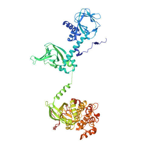
    Cyclic GMP-dependent protein kinases (PKGs) are key mediators of the nitric oxide/cyclic guanosine monophosphate (cGMP) signaling pathway that regulates biological functions as diverse as smooth muscle contraction, cardiac function, and axon guidance. Understanding how cGMP differentially triggers mammalian PKG isoforms could lead to new therapeutics that inhibit or activate PKGs, complementing drugs that target nitric oxide synthases and cyclic nucleotide phosphodiesterases in this signaling axis. Alternate splicing of PRKG1 transcripts confers distinct leucine zippers, linkers, and auto-inhibitory (AI) pseudo-substrate sequences to PKG Iα and Iβ that result in isoform-specific activation properties, but the mechanism of enzyme auto-inhibition and its alleviation by cGMP is not well understood. Here, we present a crystal structure of PKG Iβ in which the AI sequence and the cyclic nucleotide-binding (CNB) domains are bound to the catalytic domain, providing a snapshot of the auto-inhibited state. Specific contacts between the PKG Iβ AI sequence and the enzyme active site help explain isoform-specific activation constants and the effects of phosphorylation in the linker. We also present a crystal structure of a PKG I CNB domain with an activating mutation linked to Thoracic Aortic Aneurysms and Dissections. Similarity of this structure to wildtype cGMP-bound domains and differences with the auto-inhibited enzyme provide a mechanistic basis for constitutive activation. We show that PKG Iβ auto-inhibition is mediated by contacts within each monomer of the native full-length dimeric protein, and using the available structural and biochemical data we develop a model for the regulation and cooperative activation of PKGs.


  • Organizational Affiliation
    • Department of Pharmacology and Chemical Biology and Center for Drug Discovery, Baylor College of Medicine, Houston, United States.

Macromolecule Content 

  • Total Structure Weight: 141.14 kDa 
  • Atom Count: 9,432 
  • Modeled Residue Count: 1,195 
  • Deposited Residue Count: 1,234 
  • Unique protein chains: 1

Macromolecules

Find similar proteins by:|  3D Structure
Entity ID: 1
MoleculeChains  Sequence LengthOrganismDetailsImage
Isoform Beta of cGMP-dependent protein kinase 1
A, B
617Homo sapiensMutation(s): 0 
Gene Names: PRKG1PRKG1BPRKGR1APRKGR1B
EC: 2.7.11.12
UniProt & NIH Common Fund Data Resources
Find proteins for Q13976 (Homo sapiens)
Explore Q13976 
Go to UniProtKB:  Q13976
PHAROS:  Q13976
GTEx:  ENSG00000185532 
Entity Groups
Sequence Clusters30% Identity50% Identity70% Identity90% Identity95% Identity100% Identity
UniProt GroupQ13976
Sequence Annotations
Expand
Reference Sequence

Small Molecules

Ligands 3 Unique
IDChains Name / Formula / InChI Key2D Diagram3D Interactions
ANP
(Subject of Investigation/LOI)

Query on ANP


Download:Ideal Coordinates CCD File
E [auth A],
I [auth B]
PHOSPHOAMINOPHOSPHONIC ACID-ADENYLATE ESTER
C10 H17 N6 O12 P3
PVKSNHVPLWYQGJ-KQYNXXCUSA-N
EDO

Query on EDO


Download:Ideal Coordinates CCD File
F [auth A],
J [auth B],
K [auth B]
1,2-ETHANEDIOL
C2 H6 O2
LYCAIKOWRPUZTN-UHFFFAOYSA-N
MN

Query on MN


Download:Ideal Coordinates CCD File
C [auth A],
D [auth A],
G [auth B],
H [auth B]
MANGANESE (II) ION
Mn
WAEMQWOKJMHJLA-UHFFFAOYSA-N
Modified Residues  1 Unique
IDChains TypeFormula2D DiagramParent
TPO
Query on TPO
A, B
L-PEPTIDE LINKINGC4 H10 N O6 PTHR

Experimental Data & Validation

Experimental Data

  • Method: X-RAY DIFFRACTION
  • Resolution: 2.41 Å
  • R-Value Free:  0.238 (Depositor), 0.243 (DCC) 
  • R-Value Work:  0.200 (Depositor), 0.205 (DCC) 
  • R-Value Observed: 0.201 (Depositor) 
Space Group: P 21 21 21
Unit Cell:
Length ( Å )Angle ( ˚ )
a = 99.832α = 90
b = 112.824β = 90
c = 150.72γ = 90
Software Package:
Software NamePurpose
PHENIXrefinement
MOSFLMdata reduction
Aimlessdata scaling
PHASERphasing

Structure Validation

View Full Validation Report


Ligand Structure Quality Assessment 


Entry History 

& Funding Information

Deposition Data


Funding OrganizationLocationGrant Number
National Institutes of Health/National Institute of General Medical Sciences (NIH/NIGMS)United StatesGM090161

Revision History  (Full details and data files)

  • Version 1.0: 2022-08-24
    Type: Initial release
  • Version 1.1: 2023-10-18
    Changes: Data collection, Refinement description
  • Version 1.2: 2024-11-20
    Changes: Structure summary