7LTM | pdb_00007ltm

Hum8 capsid


Experimental Data Snapshot

  • Method: ELECTRON MICROSCOPY
  • Resolution: 2.49 Å
  • Aggregation State: PARTICLE 
  • Reconstruction Method: SINGLE PARTICLE 

wwPDB Validation 3D Report Full Report

Validation slider image for 7LTM

This is version 2.1 of the entry. See complete history

Literature

Receptor Switching in Newly Evolved Adeno-associated Viruses.

Havlik, L.P.Das, A.Mietzsch, M.Oh, D.K.Ark, J.McKenna, R.Agbandje-McKenna, M.Asokan, A.

(2021) J Virol 95: e0058721-e0058721

  • DOI: https://doi.org/10.1128/JVI.00587-21
  • Primary Citation Related Structures: 
    7LTM

  • PubMed Abstract: 

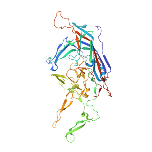
    Adeno-associated viruses utilize different glycans and the AAV receptor (AAVR) for cellular attachment and entry. Directed evolution has yielded new AAV variants; however, structure-function correlates underlying their improved transduction are generally overlooked. Here, we report that infectious cycling of structurally diverse AAV surface loop libraries yields functionally distinct variants. Newly evolved variants show enhanced cellular binding, uptake, and transduction, but through distinct mechanisms. Using glycan-based and genome-wide CRISPR knockout screens, we discover that one AAV variant acquires the ability to recognize sulfated glycosaminoglycans, while another displays receptor switching from AAVR to integrin β1 (ITGB1). A previously evolved variant, AAVhum.8, preferentially utilizes the ITGB1 receptor over AAVR. Visualization of the AAVhum.8 capsid by cryoelectron microscopy at 2.49-Å resolution localizes the newly acquired integrin recognition motif adjacent to the AAVR footprint. These observations underscore the new finding that distinct AAV surface epitopes can be evolved to exploit different cellular receptors for enhanced transduction. IMPORTANCE Understanding how viruses interact with host cells through cell surface receptors is central to discovery and development of antiviral therapeutics, vaccines, and gene transfer vectors. Here, we demonstrate that distinct epitopes on the surface of adeno-associated viruses can be evolved by infectious cycling to recognize different cell surface carbohydrates and glycoprotein receptors and solve the three-dimensional structure of one such newly evolved AAV capsid, which provides a roadmap for designing viruses with improved attributes for gene therapy applications.


  • Organizational Affiliation
    • Curriculum in Genetics and Molecular Biology, University of North Carolina at Chapel Hillgrid.10698.36, Chapel Hill, North Carolina, USA.

Macromolecule Content 

  • Total Structure Weight: 3,529.36 kDa 
  • Atom Count: 249,300 
  • Modeled Residue Count: 31,080 
  • Deposited Residue Count: 31,080 
  • Unique protein chains: 1

Macromolecules

Find similar proteins by:|  3D Structure
Entity ID: 1
MoleculeChains  Sequence LengthOrganismDetailsImage
Capsid protein518adeno-associated virus 8Mutation(s): 0 
UniProt
Find proteins for Q8JQF8 (Adeno-associated virus 8)
Explore Q8JQF8 
Go to UniProtKB:  Q8JQF8
Entity Groups
Sequence Clusters30% Identity50% Identity70% Identity90% Identity95% Identity100% Identity
UniProt GroupQ8JQF8
Sequence Annotations
Expand
Reference Sequence

Small Molecules

Ligands 1 Unique
IDChains Name / Formula / InChI Key2D Diagram3D Interactions
D5M

Query on D5M


Download:Ideal Coordinates CCD File
AC [auth S]
AD [auth s]
BC [auth T]
BD [auth t]
CC [auth U]
AC [auth S],
AD [auth s],
BC [auth T],
BD [auth t],
CC [auth U],
CD [auth u],
DC [auth V],
DD [auth v],
EC [auth W],
ED [auth w],
FC [auth X],
FD [auth x],
GC [auth Y],
GD [auth y],
HC [auth Z],
HD [auth z],
IB [auth A],
IC [auth a],
ID [auth 1],
JB [auth B],
JC [auth b],
JD [auth 2],
KB [auth C],
KC [auth c],
KD [auth 3],
LB [auth D],
LC [auth d],
LD [auth 4],
MB [auth E],
MC [auth e],
MD [auth 5],
NB [auth F],
NC [auth f],
ND [auth 6],
OB [auth G],
OC [auth g],
OD [auth 7],
PB [auth H],
PC [auth h],
PD [auth 8],
QB [auth I],
QC [auth i],
RB [auth J],
RC [auth j],
SB [auth K],
SC [auth k],
TB [auth L],
TC [auth l],
UB [auth M],
UC [auth m],
VB [auth N],
VC [auth n],
WB [auth O],
WC [auth o],
XB [auth P],
XC [auth p],
YB [auth Q],
YC [auth q],
ZB [auth R],
ZC [auth r]
2'-DEOXYADENOSINE-5'-MONOPHOSPHATE
C10 H14 N5 O6 P
KHWCHTKSEGGWEX-RRKCRQDMSA-N

Experimental Data & Validation

Experimental Data

  • Method: ELECTRON MICROSCOPY
  • Resolution: 2.49 Å
  • Aggregation State: PARTICLE 
  • Reconstruction Method: SINGLE PARTICLE 
EM Software:
TaskSoftware PackageVersion
MODEL REFINEMENTPHENIX
RECONSTRUCTIONcisTEM

Structure Validation

View Full Validation Report


Entry History 

& Funding Information

Deposition Data


Funding OrganizationLocationGrant Number
National Institutes of Health/National Institute of General Medical Sciences (NIH/NIGMS)United StatesGM082946

Revision History  (Full details and data files)

  • Version 1.0: 2021-07-07
    Type: Initial release
  • Version 1.1: 2021-08-18
    Changes: Database references
  • Version 1.2: 2021-09-22
    Changes: Database references
  • Version 2.0: 2024-05-29
    Changes: Advisory, Atomic model, Data collection, Derived calculations, Non-polymer description
  • Version 2.1: 2025-05-28
    Changes: Data collection, Structure summary