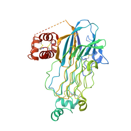
7LS0

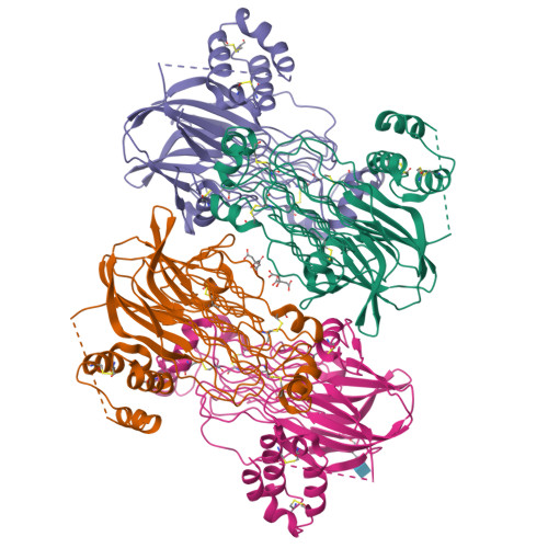
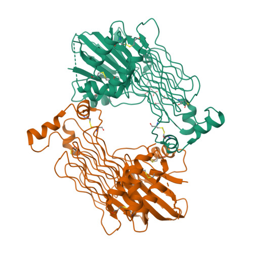
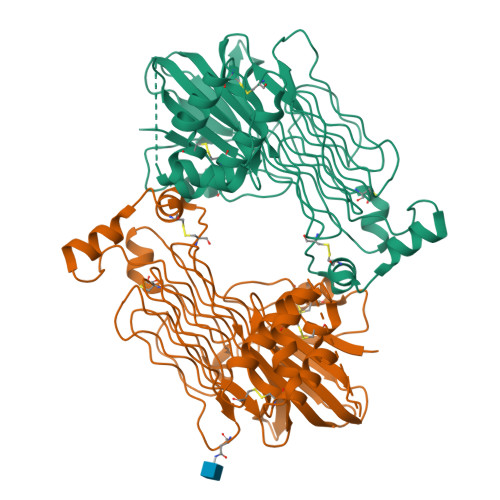
Structure of the Human ALK GRD bound to AUG


Experimental Data Snapshot

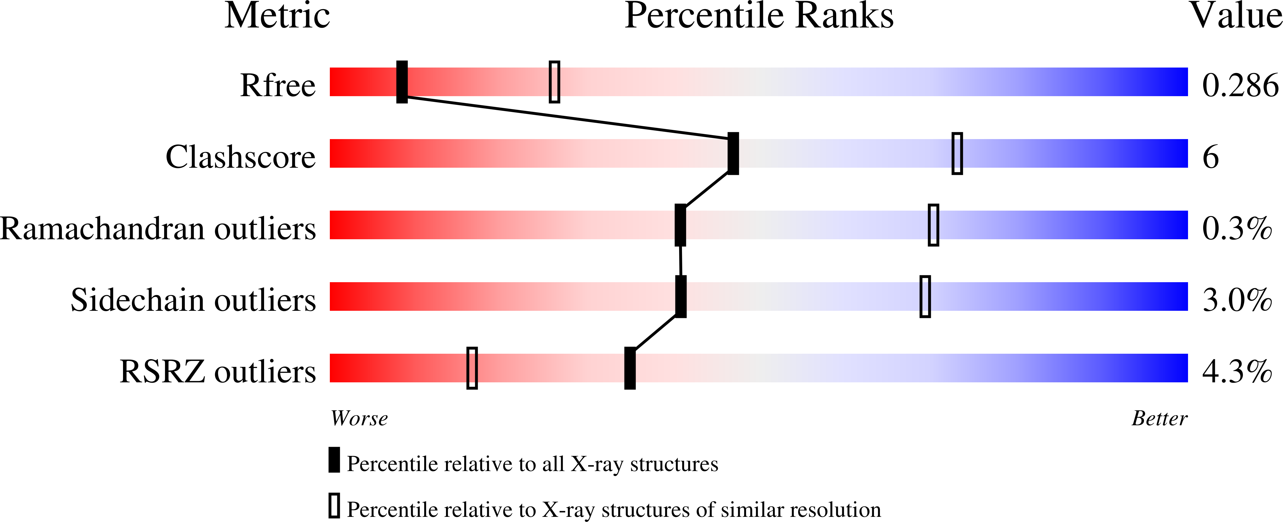
  • Method: X-RAY DIFFRACTION
  • Resolution: 3.05 Å
  • R-Value Free: 0.288 
  • R-Value Work: 0.238 
  • R-Value Observed: 0.240 

Starting Model: experimental
View more details

wwPDB Validation   3D Report Full Report


Ligand Structure Quality Assessment 


This is version 1.4 of the entry. See complete history


Literature

Structural basis for ligand reception by anaplastic lymphoma kinase.

Li, T.Stayrook, S.E.Tsutsui, Y.Zhang, J.Wang, Y.Li, H.Proffitt, A.Krimmer, S.G.Ahmed, M.Belliveau, O.Walker, I.X.Mudumbi, K.C.Suzuki, Y.Lax, I.Alvarado, D.Lemmon, M.A.Schlessinger, J.Klein, D.E.

(2021) Nature 600: 148-152

  • DOI: https://doi.org/10.1038/s41586-021-04141-7
  • Primary Citation of Related Structures:  
    7LIR, 7LRZ, 7LS0, 7MK7

  • PubMed Abstract: 

    The proto-oncogene ALK encodes anaplastic lymphoma kinase, a receptor tyrosine kinase that is expressed primarily in the developing nervous system. After development, ALK activity is associated with learning and memory 1 and controls energy expenditure, and inhibition of ALK can prevent diet-induced obesity 2 . Aberrant ALK signalling causes numerous cancers 3 . In particular, full-length ALK is an important driver in paediatric neuroblastoma 4,5 , in which it is either mutated 6 or activated by ligand 7 . Here we report crystal structures of the extracellular glycine-rich domain (GRD) of ALK, which regulates receptor activity by binding to activating peptides 8,9 . Fusing the ALK GRD to its ligand enabled us to capture a dimeric receptor complex that reveals how ALK responds to its regulatory ligands. We show that repetitive glycines in the GRD form rigid helices that separate the major ligand-binding site from a distal polyglycine extension loop (PXL) that mediates ALK dimerization. The PXL of one receptor acts as a sensor for the complex by interacting with a ligand-bound second receptor. ALK activation can be abolished through PXL mutation or with PXL-targeting antibodies. Together, these results explain how ALK uses its atypical architecture for its regulation, and suggest new therapeutic opportunities for ALK-expressing cancers such as paediatric neuroblastoma.


  • Organizational Affiliation

    Department of Pharmacology, Yale University School of Medicine, New Haven, CT, USA.


Macromolecules
Find similar proteins by:  (by identity cutoff)  |  3D Structure
Entity ID: 1
MoleculeChains Sequence LengthOrganismDetailsImage
ALK tyrosine kinase receptor fused with ALK and LTK ligand 2
A, B, C, D
451Homo sapiensMutation(s): 0 
Gene Names: ALKALKAL2FAM150BUNQ542/PRO1097
EC: 2.7.10.1
UniProt & NIH Common Fund Data Resources
Find proteins for Q6UX46 (Homo sapiens)
Explore Q6UX46 
Go to UniProtKB:  Q6UX46
PHAROS:  Q6UX46
GTEx:  ENSG00000189292 
Find proteins for Q9UM73 (Homo sapiens)
Explore Q9UM73 
Go to UniProtKB:  Q9UM73
PHAROS:  Q9UM73
GTEx:  ENSG00000171094 
Entity Groups  
Sequence Clusters30% Identity50% Identity70% Identity90% Identity95% Identity100% Identity
UniProt GroupsQ6UX46Q9UM73
Glycosylation
Glycosylation Sites: 1Go to GlyGen: Q9UM73-1Q6UX46-1
Sequence Annotations
Expand
  • Reference Sequence
Experimental Data & Validation

Experimental Data

  • Method: X-RAY DIFFRACTION
  • Resolution: 3.05 Å
  • R-Value Free: 0.288 
  • R-Value Work: 0.238 
  • R-Value Observed: 0.240 
  • Space Group: P 1 21 1
Unit Cell:
Length ( Å )Angle ( ˚ )
a = 66.262α = 90
b = 177.754β = 104.8
c = 94.855γ = 90
Software Package:
Software NamePurpose
PHENIXrefinement
XDSdata reduction
SCALAdata scaling
PDB_EXTRACTdata extraction
PHASERphasing

Structure Validation

View Full Validation Report


Ligand Structure Quality Assessment 


Entry History & Funding Information

Deposition Data


Funding OrganizationLocationGrant Number
National Institutes of Health/National Cancer Institute (NIH/NCI)United StatesK22 CA215821
National Institutes of Health/National Cancer Institute (NIH/NCI)United StatesR01 CA248532

Revision History  (Full details and data files)

  • Version 1.0: 2021-11-24
    Type: Initial release
  • Version 1.1: 2021-12-08
    Changes: Database references
  • Version 1.2: 2021-12-15
    Changes: Database references
  • Version 1.3: 2023-10-18
    Changes: Data collection, Refinement description
  • Version 1.4: 2024-11-13
    Changes: Structure summary