7LQN | pdb_00007lqn

Glucosamine-6-phosphate Deaminase from H. influenzae


Experimental Data Snapshot

  • Method: X-RAY DIFFRACTION
  • Resolution: 3.00 Å
  • R-Value Free: 
    0.242 (Depositor), 0.246 (DCC) 
  • R-Value Work: 
    0.194 (Depositor), 0.197 (DCC) 
  • R-Value Observed: 
    0.196 (Depositor) 

Starting Model: experimental
View more details

wwPDB Validation 3D Report Full Report

Validation slider image for 7LQN

This is version 1.2 of the entry. See complete history

Literature

A dimer between monomers and hexamers-Oligomeric variations in glucosamine-6-phosphate deaminase family.

Srinivasachari, S.Tiwari, V.R.Kharbanda, T.Sowdamini, R.Subramanian, R.

(2023) PLoS One 18: e0271654-e0271654

  • DOI: https://doi.org/10.1371/journal.pone.0271654
  • Primary Citation Related Structures: 
    7LQM, 7LQN

  • PubMed Abstract: 

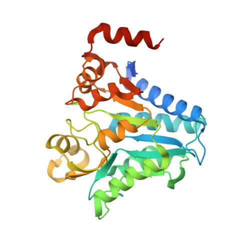
    In bacteria that live in hosts whose terminal sugar is a sialic acid, Glucosamine-6-phosphate deaminase (NagB) catalyzes the last step in converting sialic acid into Fructose-6-phosphate. These bacteria then use the Fructose-6-phosphate as an energy source. The enzyme NagB exists as a hexamer in Gram-negative bacteria and is allosterically regulated. In Gram-positive bacteria, it exists as a monomer and lacks allosteric regulation. Our identification of a dimeric Gram-negative bacterial NagB motivated us to characterize the structural basis of two closely related oligomeric forms. We report here the crystal structures of NagB from two Gram-negative pathogens, Haemophilus influenzae (Hi) and Pasturella multocida (Pm). The Hi-NagB is active as a hexamer, while Pm-NagB is active as a dimer. Both Hi-NagB and Pm-NagB contain the C-terminal helix implicated as essential for hexamer formation. The hexamer is described as a dimer of trimers. In the Pm-NagB dimer, the dimeric interface is conserved. The conservation of the dimer interface suggests that the three possible oligomeric forms of NagB are a monomer, a dimer, and a trimer of dimers. Computational modeling and MD simulations indicate that the residues at the trimeric interface have less stabilizing energy of oligomer formation than those in the dimer interface. We propose that Pm-NagB is the evolutionary link between the monomer and the hexamer forms.


  • Organizational Affiliation
    • Institute for Stem Cell Science and Regenerative Medicine, Bengaluru, Karnataka, India.

Macromolecule Content 

  • Total Structure Weight: 389.52 kDa 
  • Atom Count: 25,020 
  • Modeled Residue Count: 3,132 
  • Deposited Residue Count: 3,432 
  • Unique protein chains: 1

Macromolecules

Find similar proteins by:|  3D Structure
Entity ID: 1
MoleculeChains  Sequence LengthOrganismDetailsImage
Glucosamine-6-phosphate deaminase
A, B, C, D, E
A, B, C, D, E, F, G, H, I, J, K, L
286Haemophilus influenzae 86-028NPMutation(s): 0 
Gene Names: nagBNTHI0227
EC: 3.5.99.6
UniProt
Find proteins for Q4QP46 (Haemophilus influenzae (strain 86-028NP))
Explore Q4QP46 
Go to UniProtKB:  Q4QP46
Entity Groups
Sequence Clusters30% Identity50% Identity70% Identity90% Identity95% Identity100% Identity
UniProt GroupQ4QP46
Sequence Annotations
Expand
Reference Sequence

Experimental Data & Validation

Experimental Data

  • Method: X-RAY DIFFRACTION
  • Resolution: 3.00 Å
  • R-Value Free:  0.242 (Depositor), 0.246 (DCC) 
  • R-Value Work:  0.194 (Depositor), 0.197 (DCC) 
  • R-Value Observed: 0.196 (Depositor) 
Space Group: P 1 21 1
Unit Cell:
Length ( Å )Angle ( ˚ )
a = 103.405α = 90
b = 144.3β = 92.07
c = 131.143γ = 90
Software Package:
Software NamePurpose
XDSdata reduction
Aimlessdata scaling
PHENIXrefinement
PDB_EXTRACTdata extraction
PHENIXphasing

Structure Validation

View Full Validation Report


Entry History 

& Funding Information

Deposition Data


Funding OrganizationLocationGrant Number
Department of Biotechnology (DBT, India)IndiaBT/IN/Sweden/06/SR/2017-18
Wellcome TrustUnited Kingdom500210-Z-11-Z

Revision History  (Full details and data files)

  • Version 1.0: 2022-01-19
    Type: Initial release
  • Version 1.1: 2023-08-02
    Changes: Author supporting evidence, Database references, Refinement description
  • Version 1.2: 2023-10-18
    Changes: Data collection, Refinement description