7LIA | pdb_00007lia

5-HT bound serotonin transporter reconstituted in lipid nanodisc in presence of NaCl in outward facing conformation


Experimental Data Snapshot

  • Method: ELECTRON MICROSCOPY
  • Resolution: 3.30 Å
  • Aggregation State: PARTICLE 
  • Reconstruction Method: SINGLE PARTICLE 

wwPDB Validation 3D Report Full Report

Validation slider image for 7LIA

This is version 1.2 of the entry. See complete history

Literature

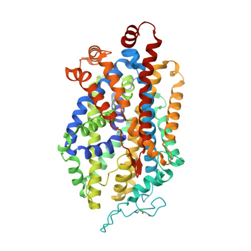
Illumination of serotonin transporter mechanism and role of the allosteric site.

Yang, D.Gouaux, E.

(2021) Sci Adv 7: eabl3857-eabl3857

  • DOI: https://doi.org/10.1126/sciadv.abl3857
  • Primary Citation Related Structures: 
    7LI6, 7LI7, 7LI8, 7LI9, 7LIA, 7MGW

  • PubMed Abstract: 

    The serotonin transporter (SERT) terminates serotonin signaling by using sodium and chloride gradients to drive reuptake of serotonin into presynaptic neurons and is the target of widely used medications to treat neuropsychiatric disorders. Despite decades of study, the molecular mechanism of serotonin transport, the coupling to ion gradients, and the role of the allosteric site have remained elusive. Here, we present cryo–electron microscopy structures of SERT in serotonin-bound and serotonin-free states, in the presence of sodium or potassium, resolving all fundamental states of the transport cycle. From the SERT-serotonin complex, we localize the substrate-bound allosteric site, formed by an aromatic pocket positioned in the scaffold domain in the extracellular vestibule, connected to the central site via a short tunnel. Together with elucidation of multiple apo state conformations, we provide previously unseen structural understanding of allosteric modulation, demonstrating how SERT binds serotonin from synaptic volumes and promotes unbinding into the presynaptic neurons.


  • Organizational Affiliation
    • Vollum Institute, Oregon Health and Science University, Portland, OR 97239, USA.

Macromolecule Content 

  • Total Structure Weight: 88.96 kDa 
  • Atom Count: 6,279 
  • Modeled Residue Count: 767 
  • Deposited Residue Count: 767 
  • Unique protein chains: 3

Macromolecules

Find similar proteins by:|  3D Structure
Entity ID: 1
MoleculeChains  Sequence LengthOrganismDetailsImage
Sodium-dependent serotonin transporter539Homo sapiensMutation(s): 0 
Gene Names: SLC6A4HTTSERT
Membrane Entity: Yes 
UniProt & NIH Common Fund Data Resources
Find proteins for P31645 (Homo sapiens)
Explore P31645 
Go to UniProtKB:  P31645
PHAROS:  P31645
GTEx:  ENSG00000108576 
Entity Groups
Sequence Clusters30% Identity50% Identity70% Identity90% Identity95% Identity100% Identity
UniProt GroupP31645
Glycosylation
Glycosylation Sites: 1Go to GlyGen: P31645-1
Sequence Annotations
Expand
Reference Sequence
Find similar proteins by:|  3D Structure
Entity ID: 2
MoleculeChains  Sequence LengthOrganismDetailsImage
variable domain of 15B8 antibody Fab heavy chain118Mus musculusMutation(s): 0 
Find similar proteins by:|  3D Structure
Entity ID: 3
MoleculeChains  Sequence LengthOrganismDetailsImage
variable domain of 15B8 antibody Fab light chain110Mus musculusMutation(s): 0 

Oligosaccharides

Help  
Entity ID: 4
MoleculeChains Length2D Diagram GlycosylationD Interactions
2-acetamido-2-deoxy-beta-D-glucopyranose-(1-4)-2-acetamido-2-deoxy-beta-D-glucopyranose
D
2N-Glycosylation
Glycosylation Resources
GlyTouCan: G42666HT
GlyCosmos: G42666HT
GlyGen: G42666HT

Small Molecules

Ligands 7 Unique
IDChains Name / Formula / InChI Key2D Diagram3D Interactions
CLR

Query on CLR


Download:Ideal Coordinates CCD File
CA [auth A]CHOLESTEROL
C27 H46 O
HVYWMOMLDIMFJA-DPAQBDIFSA-N
SRO
(Subject of Investigation/LOI)

Query on SRO


Download:Ideal Coordinates CCD File
E [auth A],
I [auth A]
SEROTONIN
C10 H12 N2 O
QZAYGJVTTNCVMB-UHFFFAOYSA-N
D10

Query on D10


Download:Ideal Coordinates CCD File
AA [auth A],
BA [auth A],
J [auth A],
V [auth A],
W [auth A]
DECANE
C10 H22
DIOQZVSQGTUSAI-UHFFFAOYSA-N
HP6

Query on HP6


Download:Ideal Coordinates CCD File
K [auth A]
L [auth A]
M [auth A]
N [auth A]
O [auth A]
K [auth A],
L [auth A],
M [auth A],
N [auth A],
O [auth A],
P [auth A],
Q [auth A],
R [auth A],
S [auth A],
T [auth A],
X [auth A],
Y [auth A],
Z [auth A]
HEPTANE
C7 H16
IMNFDUFMRHMDMM-UHFFFAOYSA-N
LNK

Query on LNK


Download:Ideal Coordinates CCD File
U [auth A]PENTANE
C5 H12
OFBQJSOFQDEBGM-UHFFFAOYSA-N
CL

Query on CL


Download:Ideal Coordinates CCD File
F [auth A]CHLORIDE ION
Cl
VEXZGXHMUGYJMC-UHFFFAOYSA-M
NA

Query on NA


Download:Ideal Coordinates CCD File
G [auth A],
H [auth A]
SODIUM ION
Na
FKNQFGJONOIPTF-UHFFFAOYSA-N

Experimental Data & Validation

Experimental Data

  • Method: ELECTRON MICROSCOPY
  • Resolution: 3.30 Å
  • Aggregation State: PARTICLE 
  • Reconstruction Method: SINGLE PARTICLE 
EM Software:
TaskSoftware PackageVersion
MODEL REFINEMENTPHENIX

Structure Validation

View Full Validation Report


Entry History 

& Funding Information

Deposition Data


Funding OrganizationLocationGrant Number
Howard Hughes Medical Institute (HHMI)United States--
National Institutes of Health/National Institute of Mental Health (NIH/NIMH)United States--

Revision History  (Full details and data files)

  • Version 1.0: 2021-12-15
    Type: Initial release
  • Version 1.1: 2024-10-30
    Changes: Data collection, Structure summary
  • Version 1.2: 2025-05-28
    Changes: Data collection