7LF8 | pdb_00007lf8

Fab 6D12 bound to ApoL2 NTD


Experimental Data Snapshot

  • Method: X-RAY DIFFRACTION
  • Resolution: 2.15 Å
  • R-Value Free: 
    0.230 (Depositor), 0.249 (DCC) 
  • R-Value Work: 
    0.192 (Depositor), 0.213 (DCC) 
  • R-Value Observed: 
    0.194 (Depositor) 

Starting Model: experimental
View more details

wwPDB Validation 3D Report Full Report

Validation slider image for 7LF8

This is version 1.3 of the entry. See complete history

Literature

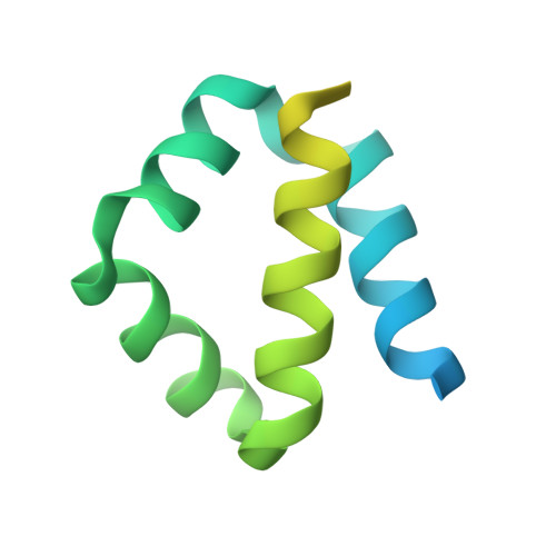
Structures of the ApoL1 and ApoL2 N-terminal domains reveal a non-classical four-helix bundle motif.

Ultsch, M.Holliday, M.J.Gerhardy, S.Moran, P.Scales, S.J.Gupta, N.Oltrabella, F.Chiu, C.Fairbrother, W.Eigenbrot, C.Kirchhofer, D.

(2021) Commun Biol 4: 916-916

  • DOI: https://doi.org/10.1038/s42003-021-02387-5
  • Primary Citation Related Structures: 
    7L6K, 7LF7, 7LF8, 7LFA, 7LFB, 7LFD

  • PubMed Abstract: 

    Apolipoprotein L1 (ApoL1) is a circulating innate immunity protein protecting against trypanosome infection. However, two ApoL1 coding variants are associated with a highly increased risk of chronic kidney disease. Here we present X-ray and NMR structures of the N-terminal domain (NTD) of ApoL1 and of its closest relative ApoL2. In both proteins, four of the five NTD helices form a four-helix core structure which is different from the classical four-helix bundle and from the pore-forming domain of colicin A. The reactivity with a conformation-specific antibody and structural models predict that this four-helix motif is also present in the NTDs of ApoL3 and ApoL4, suggesting related functions within the small ApoL family. The long helix 5 of ApoL1 is conformationally flexible and contains the BH3-like region. This BH3-like α-helix resembles true BH3 domains only in sequence and structure but not in function, since it does not bind to the pro-survival members of the Bcl-2 family, suggesting a Bcl-2-independent role in cytotoxicity. These findings should expedite a more comprehensive structural and functional understanding of the ApoL immune protein family.


  • Organizational Affiliation
    • Department of Structural Biology, Genentech Inc., South San Francisco, CA, USA.

Macromolecule Content 

  • Total Structure Weight: 63.51 kDa 
  • Atom Count: 3,831 
  • Modeled Residue Count: 486 
  • Deposited Residue Count: 567 
  • Unique protein chains: 3

Macromolecules

Find similar proteins by:|  3D Structure
Entity ID: 1
MoleculeChains  Sequence LengthOrganismDetailsImage
Apolipoprotein L2128Homo sapiensMutation(s): 0 
Gene Names: APOL2
UniProt & NIH Common Fund Data Resources
Find proteins for Q9BQE5 (Homo sapiens)
Explore Q9BQE5 
Go to UniProtKB:  Q9BQE5
PHAROS:  Q9BQE5
GTEx:  ENSG00000128335 
Entity Groups
Sequence Clusters30% Identity50% Identity70% Identity90% Identity95% Identity100% Identity
UniProt GroupQ9BQE5
Sequence Annotations
Expand
Reference Sequence
Find similar proteins by:|  3D Structure
Entity ID: 2
MoleculeChains  Sequence LengthOrganismDetailsImage
Fab 6D12 heavy chainB [auth H]225Homo sapiensMutation(s): 0 
Find similar proteins by:|  3D Structure
Entity ID: 3
MoleculeChains  Sequence LengthOrganismDetailsImage
Fab 6D12 light chainC [auth L]214Homo sapiensMutation(s): 0 

Small Molecules

Modified Residues  1 Unique
IDChains TypeFormula2D DiagramParent
TPO
Query on TPO
B [auth H]L-PEPTIDE LINKINGC4 H10 N O6 PTHR

Experimental Data & Validation

Experimental Data

  • Method: X-RAY DIFFRACTION
  • Resolution: 2.15 Å
  • R-Value Free:  0.230 (Depositor), 0.249 (DCC) 
  • R-Value Work:  0.192 (Depositor), 0.213 (DCC) 
  • R-Value Observed: 0.194 (Depositor) 
Space Group: C 2 2 21
Unit Cell:
Length ( Å )Angle ( ˚ )
a = 97.903α = 90
b = 154.29β = 90
c = 88.054γ = 90
Software Package:
Software NamePurpose
BUSTERrefinement
PDB_EXTRACTdata extraction
DIALSdata reduction
STARANISOdata scaling
PHASERphasing

Structure Validation

View Full Validation Report


Entry History 

Deposition Data

Revision History  (Full details and data files)

  • Version 1.0: 2021-08-04
    Type: Initial release
  • Version 1.1: 2022-02-16
    Changes: Database references
  • Version 1.2: 2023-10-18
    Changes: Data collection, Refinement description
  • Version 1.3: 2024-10-09
    Changes: Structure summary