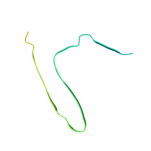
The N terminus of alpha-synuclein dictates fibril formation.
McGlinchey, R.P., Ni, X., Shadish, J.A., Jiang, J., Lee, J.C.(2021) Proc Natl Acad Sci U S A 118
- PubMed: 34452994
- DOI: https://doi.org/10.1073/pnas.2023487118
- Primary Citation of Related Structures:
7LC9 - PubMed Abstract:
The generation of α-synuclein (α-syn) truncations from incomplete proteolysis plays a significant role in the pathogenesis of Parkinson's disease. It is well established that C-terminal truncations exhibit accelerated aggregation and serve as potent seeds in fibril propagation. In contrast, mechanistic understanding of N-terminal truncations remains ill defined. Previously, we found that disease-related C-terminal truncations resulted in increased fibrillar twist, accompanied by modest conformational changes in a more compact core, suggesting that the N-terminal region could be dictating fibril structure. Here, we examined three N-terminal truncations, in which deletions of 13-, 35-, and 40-residues in the N terminus modulated both aggregation kinetics and fibril morphologies. Cross-seeding experiments showed that out of the three variants, only ΔN13-α-syn (14‒140) fibrils were capable of accelerating full-length fibril formation, albeit slower than self-seeding. Interestingly, the reversed cross-seeding reactions with full-length seeds efficiently promoted all but ΔN40-α-syn (41-140). This behavior can be explained by the unique fibril structure that is adopted by 41-140 with two asymmetric protofilaments, which was determined by cryogenic electron microscopy. One protofilament resembles the previously characterized bent β-arch kernel, comprised of residues E46‒K96, whereas in the other protofilament, fewer residues (E61‒D98) are found, adopting an extended β-hairpin conformation that does not resemble other reported structures. An interfilament interface exists between residues K60‒F94 and Q62‒I88 with an intermolecular salt bridge between K80 and E83. Together, these results demonstrate a vital role for the N-terminal residues in α-syn fibril formation and structure, offering insights into the interplay of α-syn and its truncations.
Organizational Affiliation:
Laboratory of Protein Conformation and Dynamics, Biochemistry and Biophysics Center, National Heart, Lung, and Blood Institute, Bethesda, MD 20892.


