7LA6

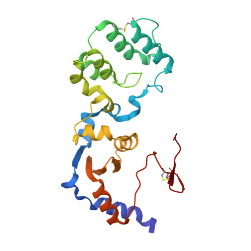
THE STRUCTURE OF A SENSOR DOMAIN OF A HISTIDINE KINASE (VxrA) FROM VIBRIO CHOLERAE O1 BIOVAR ELTOR STR. N16961, N239 deletion mutant


Experimental Data Snapshot

  • Method: X-RAY DIFFRACTION
  • Resolution: 1.98 Å
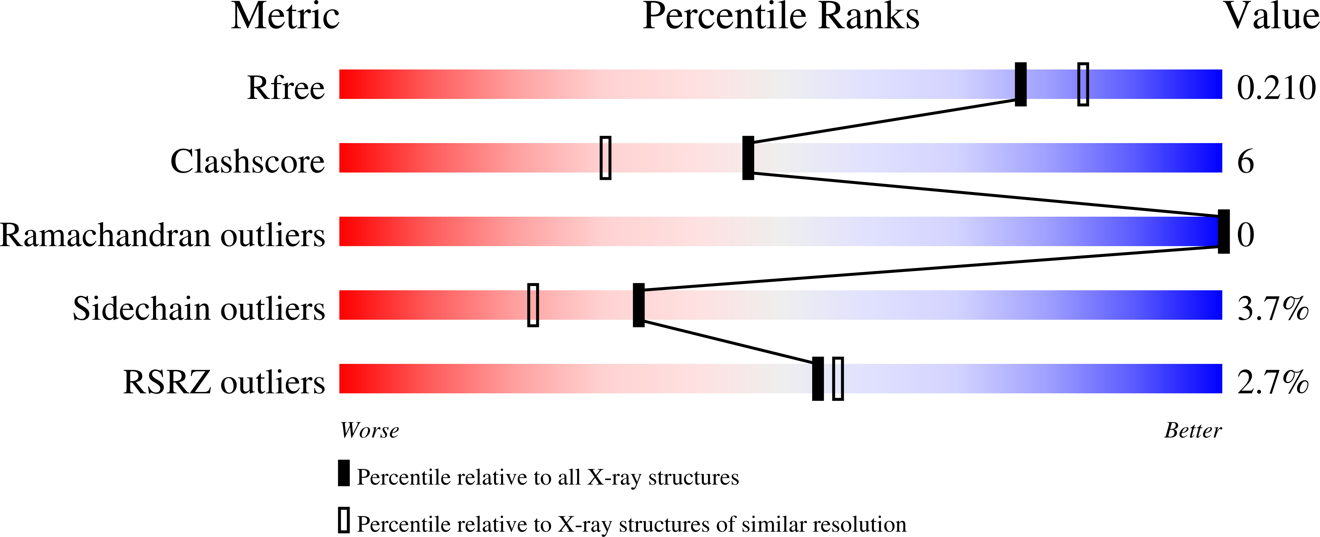
  • R-Value Free: 0.210 
  • R-Value Work: 0.181 
  • R-Value Observed: 0.183 

Starting Model: experimental
View more details

wwPDB Validation   3D Report Full Report


This is version 1.4 of the entry. See complete history


Literature

Sensor Domain of Histidine Kinase VxrA of Vibrio cholerae - A Hairpin-swapped Dimer and its Conformational Change.

Tan, K.Teschler, J.K.Wu, R.Jedrzejczak, R.P.Zhou, M.Shuvalova, L.A.Endres, M.J.Welk, L.F.Kwon, K.Anderson, W.F.Satchell, K.J.F.Yildiz, F.H.Joachimiak, A.

(2021) J Bacteriol 203

  • DOI: https://doi.org/10.1128/JB.00643-20
  • Primary Citation of Related Structures:  
    4R7Q, 7KB3, 7KB7, 7KB9, 7LA6

  • PubMed Abstract: 

    VxrA and VxrB are cognate histidine kinase (HK) - response regulator (RR) pairs of a two-component signaling system (TCS) found in Vibrio cholerae , a bacterial pathogen that causes cholera. The VxrAB TCS positively regulates virulence, the Type VI Secretion System, biofilm formation, and cell wall homeostasis in V. cholerae , providing protection from environmental stresses and contributing to the transmission and virulence of the pathogen. The VxrA HK has a unique periplasmic sensor domain (SD) and, remarkably, lacks a cytoplasmic linker domain between the second transmembrane helix and the dimerization and histidine phosphotransfer (DHp) domain, indicating that this system may utilize a potentially unique signal sensing and transmission TCS mechanism. In this study, we have determined several crystal structures of VxrA-SD and its mutants. These structures reveal a novel structural fold forming an unusual β hairpin-swapped dimer. A conformational change caused by relative rotation of the two monomers in a VxrA-SD dimer could potentially change the association of transmembrane helices and, subsequently, the pairing of cytoplasmic DHp domains. Based on the structural observation, we propose a putative scissor-like closing regulation mechanism for the VxrA HK. IMPORTANCE V. cholerae has a dynamic life cycle, which requires rapid adaptation to changing external conditions. Two-component signal transduction (TCS) systems allow V. cholerae to sense and respond to these environmental changes. The VxrAB TCS positively regulates a number of important V. cholerae phenotypes, including virulence, the Type Six Secretion System, biofilm formation, and cell wall homeostasis. Here, we provide the crystal structure of the VxrA sensor histidine kinase sensing domain and propose a mechanism for signal transduction. The cognate signal for VxrAB remains unknown, however, in this work we couple our structural analysis with functional assessments of key residues to further our understanding of this important TCS.


  • Organizational Affiliation

    Center for Structural Genomics of Infectious Diseases, University of Chicago, 5735 South Ellis Avenue, Chicago, IL 60637, USA.


Macromolecules
Find similar proteins by:  (by identity cutoff)  |  3D Structure
Entity ID: 1
MoleculeChains Sequence LengthOrganismDetailsImage
Sensor histidine kinase221Vibrio cholerae O1 biovar El Tor str. N16961Mutation(s): 0 
Gene Names: VC_A0565
EC: 2.7.13.3
UniProt
Find proteins for Q9KM24 (Vibrio cholerae serotype O1 (strain ATCC 39315 / El Tor Inaba N16961))
Explore Q9KM24 
Go to UniProtKB:  Q9KM24
Entity Groups  
Sequence Clusters30% Identity50% Identity70% Identity90% Identity95% Identity100% Identity
UniProt GroupQ9KM24
Sequence Annotations
Expand
  • Reference Sequence
Small Molecules
Ligands 4 Unique
IDChains Name / Formula / InChI Key2D Diagram3D Interactions
PG4
Query on PG4

Download Ideal Coordinates CCD File 
H [auth A]TETRAETHYLENE GLYCOL
C8 H18 O5
UWHCKJMYHZGTIT-UHFFFAOYSA-N
PEG
Query on PEG

Download Ideal Coordinates CCD File 
D [auth A],
E [auth A],
F [auth A],
G [auth A]
DI(HYDROXYETHYL)ETHER
C4 H10 O3
MTHSVFCYNBDYFN-UHFFFAOYSA-N
SO4
Query on SO4

Download Ideal Coordinates CCD File 
I [auth A],
J [auth A],
K [auth A]
SULFATE ION
O4 S
QAOWNCQODCNURD-UHFFFAOYSA-L
GOL
Query on GOL

Download Ideal Coordinates CCD File 
B [auth A],
C [auth A]
GLYCEROL
C3 H8 O3
PEDCQBHIVMGVHV-UHFFFAOYSA-N
Experimental Data & Validation

Experimental Data

  • Method: X-RAY DIFFRACTION
  • Resolution: 1.98 Å
  • R-Value Free: 0.210 
  • R-Value Work: 0.181 
  • R-Value Observed: 0.183 
  • Space Group: P 41 21 2
Unit Cell:
Length ( Å )Angle ( ˚ )
a = 87.654α = 90
b = 87.654β = 90
c = 106.223γ = 90
Software Package:
Software NamePurpose
SBC-Collectdata collection
PHENIXrefinement
HKL-3000data scaling
HKL-3000data extraction
HKL-3000phasing

Structure Validation

View Full Validation Report


Entry History & Funding Information

Deposition Data


Funding OrganizationLocationGrant Number
National Institutes of Health/National Institute Of Allergy and Infectious Diseases (NIH/NIAID)United StatesHHSN272201700060C

Revision History  (Full details and data files)

  • Version 1.0: 2021-01-27
    Type: Initial release
  • Version 1.1: 2022-11-16
    Changes: Database references
  • Version 1.2: 2022-12-07
    Changes: Database references
  • Version 1.3: 2023-10-25
    Changes: Data collection, Refinement description
  • Version 1.4: 2024-11-20
    Changes: Structure summary