7L7C | pdb_00007l7c

Crystal Structure of EcDsbA in a complex with 2-(6-(3-Methoxyphenyl)benzofuran-3-yl)acetic acid


Experimental Data Snapshot

  • Method: X-RAY DIFFRACTION
  • Resolution: 1.80 Å
  • R-Value Free: 
    0.196 (Depositor), 0.197 (DCC) 
  • R-Value Work: 
    0.166 (Depositor), 0.166 (DCC) 
  • R-Value Observed: 
    0.167 (Depositor) 

Starting Model: experimental
View more details

wwPDB Validation 3D Report Full Report

Validation slider image for 7L7C

Ligand Structure Quality Assessment 


This is version 1.3 of the entry. See complete history

Literature

Elaboration of a benzofuran scaffold and evaluation of binding affinity and inhibition of Escherichia coli DsbA: A fragment-based drug design approach to novel antivirulence compounds.

Duncan, L.F.Wang, G.Ilyichova, O.V.Dhouib, R.Totsika, M.Scanlon, M.J.Heras, B.Abbott, B.M.

(2021) Bioorg Med Chem 45: 116315-116315

  • DOI: https://doi.org/10.1016/j.bmc.2021.116315
  • Primary Citation Related Structures: 
    6XSP, 6XSQ, 6XT3, 7L76, 7L7C, 7LHP

  • PubMed Abstract: 

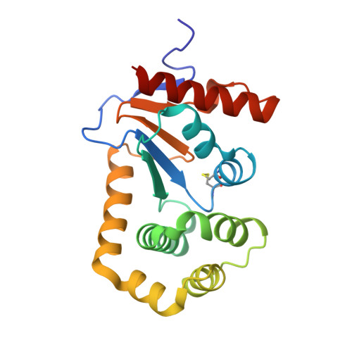
    Bacterial thiol-disulfide oxidoreductase DsbA is essential for bacterial virulence factor assembly and has been identified as a viable antivirulence target. Herein, we report a structure-based elaboration of a benzofuran hit that bound to the active site groove of Escherichia coli DsbA. Substituted phenyl groups were installed at the 5- and 6-position of the benzofuran using Suzuki-Miyaura coupling. HSQC NMR titration experiments showed dissociation constants of this series in the high µM to low mM range and X-ray crystallography produced three co-structures, showing binding in the hydrophobic groove, comparable with that of the previously reported benzofurans. The 6-(m-methoxy)phenyl analogue (2b), which showed a promising binding pose, was chosen for elaboration from the C-2 position. The 2,6-disubstituted analogues bound to the hydrophobic region of the binding groove and the C-2 groups extended into the more polar, previously un-probed, region of the binding groove. Biochemical analysis of the 2,6-disubsituted analogues showed they inhibited DsbA oxidation activity in vitro. The results indicate the potential to develop the elaborated benzofuran series into a novel class of antivirulence compounds.


  • Organizational Affiliation
    • Department of Chemistry and Physics, La Trobe Institute for Molecular Science, La Trobe University, Melbourne, VIC 3086, Australia.

Macromolecule Content 

  • Total Structure Weight: 42.66 kDa 
  • Atom Count: 3,450 
  • Modeled Residue Count: 376 
  • Deposited Residue Count: 378 
  • Unique protein chains: 1

Macromolecules

Find similar proteins by:|  3D Structure
Entity ID: 1
MoleculeChains  Sequence LengthOrganismDetailsImage
Thiol:disulfide interchange protein DsbA
A, B
189Escherichia coli K-12Mutation(s): 0 
Gene Names: dsbAdsfppfAb3860JW3832
UniProt
Find proteins for P0AEG4 (Escherichia coli (strain K12))
Explore P0AEG4 
Go to UniProtKB:  P0AEG4
Entity Groups
Sequence Clusters30% Identity50% Identity70% Identity90% Identity95% Identity100% Identity
UniProt GroupP0AEG4
Sequence Annotations
Expand
Reference Sequence

Small Molecules

Experimental Data & Validation

Experimental Data

  • Method: X-RAY DIFFRACTION
  • Resolution: 1.80 Å
  • R-Value Free:  0.196 (Depositor), 0.197 (DCC) 
  • R-Value Work:  0.166 (Depositor), 0.166 (DCC) 
  • R-Value Observed: 0.167 (Depositor) 
Space Group: C 1 2 1
Unit Cell:
Length ( Å )Angle ( ˚ )
a = 117.317α = 90
b = 63.808β = 125.023
c = 74.073γ = 90
Software Package:
Software NamePurpose
PHENIXrefinement
XDSdata reduction
SCALAdata scaling
PHASERphasing

Structure Validation

View Full Validation Report


Ligand Structure Quality Assessment 


Entry History 

Deposition Data

Revision History  (Full details and data files)

  • Version 1.0: 2021-08-11
    Type: Initial release
  • Version 1.1: 2021-08-25
    Changes: Database references
  • Version 1.2: 2023-10-18
    Changes: Data collection, Refinement description
  • Version 1.3: 2024-10-23
    Changes: Structure summary