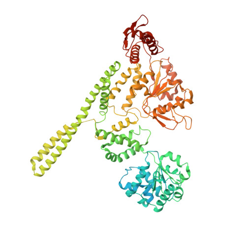
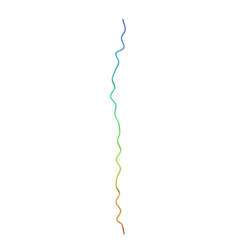
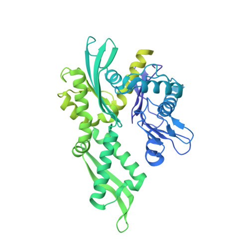
7L6N

The Mycobacterium tuberculosis ClpB disaggregase hexamer structure with three locally refined ClpB middle domains and three DnaK nucleotide binding domains


Experimental Data Snapshot

  • Method: ELECTRON MICROSCOPY
  • Resolution: 7.00 Å
  • Aggregation State: PARTICLE 
  • Reconstruction Method: SINGLE PARTICLE 

wwPDB Validation   3D Report Full Report


This is version 1.2 of the entry. See complete history


Literature

Structural basis for aggregate dissolution and refolding by the Mycobacterium tuberculosis ClpB-DnaK bi-chaperone system.

Yin, Y.Feng, X.Yu, H.Fay, A.Kovach, A.Glickman, M.S.Li, H.

(2021) Cell Rep 35: 109166-109166

  • DOI: https://doi.org/10.1016/j.celrep.2021.109166
  • Primary Citation of Related Structures:  
    6W6E, 6W6G, 6W6H, 6W6I, 6W6J, 7L6N

  • PubMed Abstract: 

    The M. tuberculosis (Mtb) ClpB is a protein disaggregase that helps to rejuvenate the bacterial cell. DnaK is a protein foldase that can function alone, but it can also bind to the ClpB hexamer to physically couple protein disaggregation with protein refolding, although the molecular mechanism is not well understood. Here, we report the cryo-EM analysis of the Mtb ClpB-DnaK bi-chaperone in the presence of ATPγS and a protein substrate. We observe three ClpB conformations in the presence of DnaK, identify a conserved TGIP loop linking the oligonucleotide/oligosaccharide-binding domain and the nucleotide-binding domain that is important for ClpB function, derive the interface between the regulatory middle domain of the ClpB and the DnaK nucleotide-binding domain, and find that DnaK binding stabilizes, but does not bend or tilt, the ClpB middle domain. We propose a model for the synergistic actions of aggregate dissolution and refolding by the Mtb ClpB-DnaK bi-chaperone system.


  • Organizational Affiliation

    Department of Structural Biology, Van Andel Institute, Grand Rapids, MI, USA.


Macromolecules
Find similar proteins by:  (by identity cutoff)  |  3D Structure
Entity ID: 1
MoleculeChains Sequence LengthOrganismDetailsImage
Chaperone protein ClpB
A, B, C, D, E
A, B, C, D, E, F
848Mycobacterium tuberculosisMutation(s): 0 
Gene Names: clpBMT0397
UniProt
Find proteins for P9WPD1 (Mycobacterium tuberculosis (strain ATCC 25618 / H37Rv))
Explore P9WPD1 
Go to UniProtKB:  P9WPD1
Entity Groups  
Sequence Clusters30% Identity50% Identity70% Identity90% Identity95% Identity100% Identity
UniProt GroupP9WPD1
Sequence Annotations
Expand
  • Reference Sequence
Find similar proteins by:  (by identity cutoff)  |  3D Structure
Entity ID: 2
MoleculeChains Sequence LengthOrganismDetailsImage
SubstrateG [auth N]33Mycobacterium tuberculosisMutation(s): 0 
Sequence Annotations
Expand
  • Reference Sequence
Find similar proteins by:  (by identity cutoff)  |  3D Structure
Entity ID: 3
MoleculeChains Sequence LengthOrganismDetailsImage
Chaperone protein DnaKH [auth I],
I [auth J],
J [auth K]
625Mycobacterium tuberculosisMutation(s): 0 
Gene Names: dnaK_2dnaKE5M05_19850ERS007703_00955ERS023446_02581ERS027651_01905FCN16_12395SAMEA2682864_01182SAMEA2683035_02658
UniProt
Find proteins for P9WMJ9 (Mycobacterium tuberculosis (strain ATCC 25618 / H37Rv))
Explore P9WMJ9 
Go to UniProtKB:  P9WMJ9
Entity Groups  
Sequence Clusters30% Identity50% Identity70% Identity90% Identity95% Identity100% Identity
UniProt GroupP9WMJ9
Sequence Annotations
Expand
  • Reference Sequence
Small Molecules
Ligands 2 Unique
IDChains Name / Formula / InChI Key2D Diagram3D Interactions
AGS (Subject of Investigation/LOI)
Query on AGS

Download Ideal Coordinates CCD File 
K [auth A]
L [auth A]
M [auth B]
N [auth B]
O [auth C]
K [auth A],
L [auth A],
M [auth B],
N [auth B],
O [auth C],
P [auth C],
Q [auth D],
R [auth D],
S [auth E],
U [auth F]
PHOSPHOTHIOPHOSPHORIC ACID-ADENYLATE ESTER
C10 H16 N5 O12 P3 S
NLTUCYMLOPLUHL-KQYNXXCUSA-N
ADP (Subject of Investigation/LOI)
Query on ADP

Download Ideal Coordinates CCD File 
T [auth E],
V [auth F]
ADENOSINE-5'-DIPHOSPHATE
C10 H15 N5 O10 P2
XTWYTFMLZFPYCI-KQYNXXCUSA-N
Experimental Data & Validation

Experimental Data

  • Method: ELECTRON MICROSCOPY
  • Resolution: 7.00 Å
  • Aggregation State: PARTICLE 
  • Reconstruction Method: SINGLE PARTICLE 

Structure Validation

View Full Validation Report


Entry History & Funding Information

Deposition Data


Funding OrganizationLocationGrant Number
National Institutes of Health/National Center for Complementary and Integrative Health (NIH/NCCIH)United StatesAI070285

Revision History  (Full details and data files)

  • Version 1.0: 2021-05-26
    Type: Initial release
  • Version 1.1: 2021-12-08
    Changes: Database references
  • Version 1.2: 2024-05-29
    Changes: Data collection