7L56 | pdb_00007l56

Cryo-EM structure of the SARS-CoV-2 spike glycoprotein bound to Fab 2-43


Experimental Data Snapshot

  • Method: ELECTRON MICROSCOPY
  • Resolution: 3.60 Å
  • Aggregation State: PARTICLE 
  • Reconstruction Method: SINGLE PARTICLE 

wwPDB Validation 3D Report Full Report

Validation slider image for 7L56

This is version 1.3 of the entry. See complete history

Literature

Modular basis for potent SARS-CoV-2 neutralization by a prevalent VH1-2-derived antibody class.

Rapp, M.Guo, Y.Reddem, E.R.Yu, J.Liu, L.Wang, P.Cerutti, G.Katsamba, P.Bimela, J.S.Bahna, F.A.Mannepalli, S.M.Zhang, B.Kwong, P.D.Huang, Y.Ho, D.D.Shapiro, L.Sheng, Z.

(2021) Cell Rep 35: 108950-108950

  • DOI: https://doi.org/10.1016/j.celrep.2021.108950
  • Primary Citation Related Structures: 
    7L56, 7L57, 7L58, 7L5B

  • PubMed Abstract: 

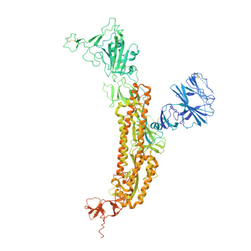
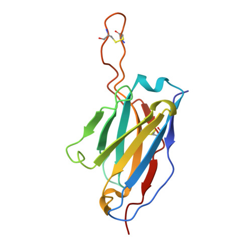
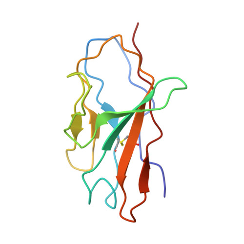
    Antibodies with heavy chains that derive from the VH1-2 gene constitute some of the most potent severe acute respiratory syndrome coronavirus 2 (SARS-CoV-2)-neutralizing antibodies yet identified. To provide insight into whether these genetic similarities inform common modes of recognition, we determine the structures of the SARS-CoV-2 spike in complex with three VH1-2-derived antibodies: 2-15, 2-43, and H4. All three use VH1-2-encoded motifs to recognize the receptor-binding domain (RBD), with heavy-chain N53I-enhancing binding and light-chain tyrosines recognizing F486 RBD . Despite these similarities, class members bind both RBD-up and -down conformations of the spike, with a subset of antibodies using elongated CDRH3s to recognize glycan N343 on a neighboring RBD-a quaternary interaction accommodated by an increase in RBD separation of up to 12 Å. The VH1-2 antibody class, thus, uses modular recognition encoded by modular genetic elements to effect potent neutralization, with the VH-gene component specifying recognition of RBD and the CDRH3 component specifying quaternary interactions.


  • Organizational Affiliation
    • Department of Biochemistry and Molecular Biophysics, Columbia University, New York, NY 10032, USA; Zuckerman Mind Brain Behavior Institute, Columbia University, New York, NY 10027, USA.

Macromolecule Content 

  • Total Structure Weight: 518.15 kDa 
  • Atom Count: 26,583 
  • Modeled Residue Count: 3,686 
  • Deposited Residue Count: 4,581 
  • Unique protein chains: 3

Macromolecules

Find similar proteins by:|  3D Structure
Entity ID: 1
MoleculeChains  Sequence LengthOrganismDetailsImage
Spike glycoprotein
A, B, C
1,288Severe acute respiratory syndrome coronavirus 2Mutation(s): 5 
Gene Names: S2
UniProt
Find proteins for P0DTC2 (Severe acute respiratory syndrome coronavirus 2)
Explore P0DTC2 
Go to UniProtKB:  P0DTC2
Entity Groups
Sequence Clusters30% Identity50% Identity70% Identity90% Identity95% Identity100% Identity
UniProt GroupP0DTC2
Glycosylation
Glycosylation Sites: 13Go to GlyGen: P0DTC2-1
Sequence Annotations
Expand
Reference Sequence
Find similar proteins by:|  3D Structure
Entity ID: 2
MoleculeChains  Sequence LengthOrganismDetailsImage
Fab 2-43 variable domain heavy chainD [auth F],
F [auth H],
G [auth J]
129Homo sapiensMutation(s): 0 
Find similar proteins by:|  3D Structure
Entity ID: 3
MoleculeChains  Sequence LengthOrganismDetailsImage
Fab 2-43 variable domain light chainE [auth G],
H [auth K],
I [auth L]
110Homo sapiensMutation(s): 0 

Oligosaccharides

Help  
Entity ID: 4
MoleculeChains Length2D Diagram GlycosylationD Interactions
2-acetamido-2-deoxy-beta-D-glucopyranose-(1-4)-2-acetamido-2-deoxy-beta-D-glucopyranose
J [auth D],
K [auth E],
N,
O,
Q,
J [auth D],
K [auth E],
N,
O,
Q,
R
2N-Glycosylation
Glycosylation Resources
GlyTouCan: G42666HT
GlyCosmos: G42666HT
GlyGen: G42666HT
Entity ID: 5
MoleculeChains Length2D Diagram GlycosylationD Interactions
beta-D-galactopyranose-(1-4)-2-acetamido-2-deoxy-beta-D-glucopyranose-(1-2)-alpha-D-mannopyranose-(1-3)-[alpha-D-mannopyranose-(1-3)-alpha-D-mannopyranose-(1-6)]beta-D-mannopyranose-(1-4)-2-acetamido-2-deoxy-beta-D-glucopyranose-(1-4)-2-acetamido-2-deoxy-beta-D-glucopyranoseL [auth I]8N-Glycosylation
Glycosylation Resources
GlyTouCan: G51367TM
GlyCosmos: G51367TM
GlyGen: G51367TM
Entity ID: 6
MoleculeChains Length2D Diagram GlycosylationD Interactions
beta-D-galactopyranose-(1-4)-2-acetamido-2-deoxy-beta-D-glucopyranose-(1-2)-alpha-D-mannopyranose-(1-3)-[alpha-D-mannopyranose-(1-6)-alpha-D-mannopyranose-(1-6)]beta-D-mannopyranose-(1-4)-2-acetamido-2-deoxy-beta-D-glucopyranose-(1-4)-2-acetamido-2-deoxy-beta-D-glucopyranose
M
8N-Glycosylation
Glycosylation Resources
GlyTouCan: G80861DM
GlyCosmos: G80861DM
GlyGen: G80861DM
Entity ID: 7
MoleculeChains Length2D Diagram GlycosylationD Interactions
beta-D-galactopyranose-(1-4)-2-acetamido-2-deoxy-beta-D-glucopyranose-(1-2)-alpha-D-mannopyranose-(1-6)-[alpha-D-mannopyranose-(1-6)-alpha-D-mannopyranose-(1-3)]beta-D-mannopyranose-(1-4)-2-acetamido-2-deoxy-beta-D-glucopyranose-(1-4)-2-acetamido-2-deoxy-beta-D-glucopyranose
P
8N-Glycosylation
Glycosylation Resources
GlyTouCan: G66227UG
GlyCosmos: G66227UG
GlyGen: G66227UG

Small Molecules

Ligands 1 Unique
IDChains Name / Formula / InChI Key2D Diagram3D Interactions
NAG

Query on NAG


Download:Ideal Coordinates CCD File
AA [auth A]
BA [auth A]
CA [auth B]
DA [auth B]
EA [auth B]
AA [auth A],
BA [auth A],
CA [auth B],
DA [auth B],
EA [auth B],
FA [auth B],
GA [auth B],
HA [auth B],
IA [auth B],
JA [auth B],
KA [auth B],
LA [auth B],
MA [auth C],
NA [auth C],
OA [auth C],
PA [auth C],
QA [auth C],
RA [auth C],
S [auth A],
SA [auth C],
T [auth A],
TA [auth C],
U [auth A],
UA [auth C],
V [auth A],
VA [auth C],
W [auth A],
WA [auth C],
X [auth A],
Y [auth A],
Z [auth A]
2-acetamido-2-deoxy-beta-D-glucopyranose
C8 H15 N O6
OVRNDRQMDRJTHS-FMDGEEDCSA-N

Experimental Data & Validation

Experimental Data

  • Method: ELECTRON MICROSCOPY
  • Resolution: 3.60 Å
  • Aggregation State: PARTICLE 
  • Reconstruction Method: SINGLE PARTICLE 

Structure Validation

View Full Validation Report


Entry History 

Deposition Data

Revision History  (Full details and data files)

  • Version 1.0: 2021-04-14
    Type: Initial release
  • Version 1.1: 2021-04-21
    Changes: Database references
  • Version 1.2: 2023-04-05
    Changes: Database references
  • Version 1.3: 2024-11-20
    Changes: Data collection, Structure summary