7L29

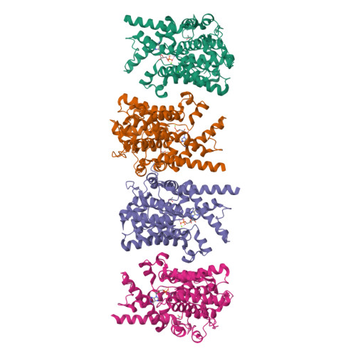
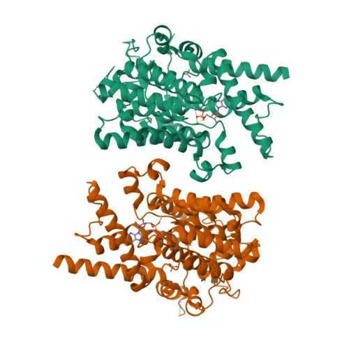
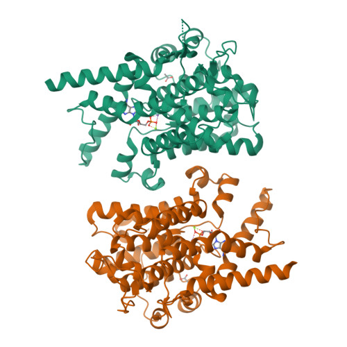
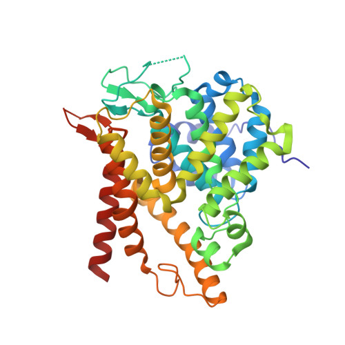
Crystal structure of the catalytic domain of human PDE3A bound to AMP


Experimental Data Snapshot

  • Method: X-RAY DIFFRACTION
  • Resolution: 2.08 Å
  • R-Value Free: 0.253 
  • R-Value Work: 0.235 
  • R-Value Observed: 0.236 

Starting Model: experimental
View more details

wwPDB Validation   3D Report Full Report


Ligand Structure Quality Assessment 


This is version 1.3 of the entry. See complete history


Literature

Structure of PDE3A-SLFN12 complex reveals requirements for activation of SLFN12 RNase.

Garvie, C.W.Wu, X.Papanastasiou, M.Lee, S.Fuller, J.Schnitzler, G.R.Horner, S.W.Baker, A.Zhang, T.Mullahoo, J.P.Westlake, L.Hoyt, S.H.Toetzl, M.Ranaghan, M.J.de Waal, L.McGaunn, J.Kaplan, B.Piccioni, F.Yang, X.Lange, M.Tersteegen, A.Raymond, D.Lewis, T.A.Carr, S.A.Cherniack, A.D.Lemke, C.T.Meyerson, M.Greulich, H.

(2021) Nat Commun 12: 4375-4375

  • DOI: https://doi.org/10.1038/s41467-021-24495-w
  • Primary Citation of Related Structures:  
    7KWE, 7L27, 7L28, 7L29, 7LRC, 7LRD, 7LRE

  • PubMed Abstract: 

    DNMDP and related compounds, or velcrins, induce complex formation between the phosphodiesterase PDE3A and the SLFN12 protein, leading to a cytotoxic response in cancer cells that express elevated levels of both proteins. The mechanisms by which velcrins induce complex formation, and how the PDE3A-SLFN12 complex causes cancer cell death, are not fully understood. Here, we show that PDE3A and SLFN12 form a heterotetramer stabilized by binding of DNMDP. Interactions between the C-terminal alpha helix of SLFN12 and residues near the active site of PDE3A are required for complex formation, and are further stabilized by interactions between SLFN12 and DNMDP. Moreover, we demonstrate that SLFN12 is an RNase, that PDE3A binding increases SLFN12 RNase activity, and that SLFN12 RNase activity is required for DNMDP response. This new mechanistic understanding will facilitate development of velcrin compounds into new cancer therapies.


  • Organizational Affiliation

    Center for the Development of Therapeutics, Broad Institute of MIT and Harvard, Cambridge, MA, USA.


Macromolecules
Find similar proteins by:  (by identity cutoff)  |  3D Structure
Entity ID: 1
MoleculeChains Sequence LengthOrganismDetailsImage
cGMP-inhibited 3',5'-cyclic phosphodiesterase A
A, B, C, D
380Homo sapiensMutation(s): 0 
Gene Names: PDE3A
EC: 3.1.4.17
UniProt & NIH Common Fund Data Resources
Find proteins for Q14432 (Homo sapiens)
Explore Q14432 
Go to UniProtKB:  Q14432
PHAROS:  Q14432
GTEx:  ENSG00000172572 
Entity Groups  
Sequence Clusters30% Identity50% Identity70% Identity90% Identity95% Identity100% Identity
UniProt GroupQ14432
Sequence Annotations
Expand
  • Reference Sequence
Small Molecules
Ligands 5 Unique
IDChains Name / Formula / InChI Key2D Diagram3D Interactions
AMP (Subject of Investigation/LOI)
Query on AMP

Download Ideal Coordinates CCD File 
E [auth A],
J [auth B],
N [auth C],
R [auth D]
ADENOSINE MONOPHOSPHATE
C10 H14 N5 O7 P
UDMBCSSLTHHNCD-KQYNXXCUSA-N
ACT
Query on ACT

Download Ideal Coordinates CCD File 
I [auth A],
M [auth B],
Q [auth C],
U [auth D]
ACETATE ION
C2 H3 O2
QTBSBXVTEAMEQO-UHFFFAOYSA-M
MN
Query on MN

Download Ideal Coordinates CCD File 
F [auth A],
K [auth B],
O [auth C],
S [auth D]
MANGANESE (II) ION
Mn
WAEMQWOKJMHJLA-UHFFFAOYSA-N
CA
Query on CA

Download Ideal Coordinates CCD File 
H [auth A]CALCIUM ION
Ca
BHPQYMZQTOCNFJ-UHFFFAOYSA-N
MG
Query on MG

Download Ideal Coordinates CCD File 
G [auth A],
L [auth B],
P [auth C],
T [auth D]
MAGNESIUM ION
Mg
JLVVSXFLKOJNIY-UHFFFAOYSA-N
Experimental Data & Validation

Experimental Data

  • Method: X-RAY DIFFRACTION
  • Resolution: 2.08 Å
  • R-Value Free: 0.253 
  • R-Value Work: 0.235 
  • R-Value Observed: 0.236 
  • Space Group: P 1 21 1
Unit Cell:
Length ( Å )Angle ( ˚ )
a = 82.415α = 90
b = 58.783β = 90.74
c = 156.996γ = 90
Software Package:
Software NamePurpose
XDSdata reduction
XSCALEdata scaling
REFMACrefinement
PDB_EXTRACTdata extraction
PHENIXphasing

Structure Validation

View Full Validation Report


Ligand Structure Quality Assessment 


Entry History 

Deposition Data

Revision History  (Full details and data files)

  • Version 1.0: 2021-06-16
    Type: Initial release
  • Version 1.1: 2021-07-28
    Changes: Database references
  • Version 1.2: 2024-03-06
    Changes: Data collection, Database references, Refinement description
  • Version 1.3: 2024-04-03
    Changes: Refinement description