7L21 | pdb_00007l21

Pyruvate Kinase M2 mutant-N70D


Experimental Data Snapshot

  • Method: X-RAY DIFFRACTION
  • Resolution: 2.29 Å
  • R-Value Free: 
    0.229 (Depositor), 0.227 (DCC) 
  • R-Value Work: 
    0.183 (Depositor), 0.184 (DCC) 
  • R-Value Observed: 
    0.186 (Depositor) 

Starting Model: experimental
View more details

wwPDB Validation 3D Report Full Report

Validation slider image for 7L21

Ligand Structure Quality Assessment 


This is version 1.2 of the entry. See complete history

Literature

Identification of residues involved in allosteric signal transmission from amino acid binding site of pyruvate kinase muscle isoform 2.

Nandi, S.Dey, M.

(2023) PLoS One 18: e0282508-e0282508

  • DOI: https://doi.org/10.1371/journal.pone.0282508
  • Primary Citation Related Structures: 
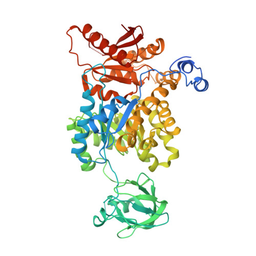
    7L21

  • PubMed Abstract: 

    PKM2 is a rate-limiting enzyme in the glycolytic process and is involved in regulating tumor proliferation. Several amino acids (AAs) such as Asn, Asp, Val, and Cys have been shown to bind to the AA binding pocket of PKM2 and modulate its oligomeric state, substrate binding affinity, and activity. Although previous studies have attributed that the main chain and side chain of bound AAs are responsible for initiating signal to regulate PKM2, the signal transduction pathway remains elusive. To identify the residues involved in signal transfer process, N70 and N75 located at two ends of a β strand connecting the active site and AA binding pocket were altered. Biochemical studies of these variants with various AA ligands (Asn, Asp, Val, and Cys), illustrate that N70 and N75, along with β1 connecting these residues are part of the signal transduction pathway between the AA binding pocket and the active site. The results demonstrate that mutation of N70 to D prevents the transfer of the inhibitory signal mediated by Val and Cys, whereas N75 to L alteration blocks the activating signal initiated by Asn and Asp. Taken together, this study confirms that N70 is one of the residues responsible for transmitting the inhibitory signal and N75 is involved in the activation signal flow.


  • Organizational Affiliation
    • Department of Chemistry, The University of Iowa, Iowa City, IA, United States of America.

Macromolecule Content 

  • Total Structure Weight: 243.03 kDa 
  • Atom Count: 16,070 
  • Modeled Residue Count: 2,052 
  • Deposited Residue Count: 2,200 
  • Unique protein chains: 1

Macromolecules

Find similar proteins by:|  3D Structure
Entity ID: 1
MoleculeChains  Sequence LengthOrganismDetailsImage
Pyruvate kinase PKM
A, B, C, D
550Homo sapiensMutation(s): 1 
Gene Names: PKMOIP3PK2PK3PKM2
EC: 2.7.1.40 (PDB Primary Data), 2.7.11.1 (UniProt), 2.7.10.2 (UniProt)
UniProt & NIH Common Fund Data Resources
Find proteins for P14618 (Homo sapiens)
Explore P14618 
Go to UniProtKB:  P14618
PHAROS:  P14618
GTEx:  ENSG00000067225 
Entity Groups
Sequence Clusters30% Identity50% Identity70% Identity90% Identity95% Identity100% Identity
UniProt GroupP14618
Sequence Annotations
Expand
Reference Sequence

Small Molecules

Ligands 4 Unique
IDChains Name / Formula / InChI Key2D Diagram3D Interactions
FBP

Query on FBP


Download:Ideal Coordinates CCD File
AA [auth D],
I [auth A],
O [auth B],
U [auth C]
1,6-di-O-phosphono-beta-D-fructofuranose
C6 H14 O12 P2
RNBGYGVWRKECFJ-ARQDHWQXSA-N
GOL

Query on GOL


Download:Ideal Coordinates CCD File
G [auth A]
H [auth A]
L [auth B]
M [auth B]
N [auth B]
G [auth A],
H [auth A],
L [auth B],
M [auth B],
N [auth B],
R [auth C],
S [auth C],
T [auth C],
X [auth D],
Y [auth D],
Z [auth D]
GLYCEROL
C3 H8 O3
PEDCQBHIVMGVHV-UHFFFAOYSA-N
OXL

Query on OXL


Download:Ideal Coordinates CCD File
E [auth A],
J [auth B],
P [auth C],
V [auth D]
OXALATE ION
C2 O4
MUBZPKHOEPUJKR-UHFFFAOYSA-L
MG

Query on MG


Download:Ideal Coordinates CCD File
F [auth A],
K [auth B],
Q [auth C],
W [auth D]
MAGNESIUM ION
Mg
JLVVSXFLKOJNIY-UHFFFAOYSA-N

Experimental Data & Validation

Experimental Data

  • Method: X-RAY DIFFRACTION
  • Resolution: 2.29 Å
  • R-Value Free:  0.229 (Depositor), 0.227 (DCC) 
  • R-Value Work:  0.183 (Depositor), 0.184 (DCC) 
  • R-Value Observed: 0.186 (Depositor) 
Space Group: C 2 2 21
Unit Cell:
Length ( Å )Angle ( ˚ )
a = 139.336α = 90
b = 158.303β = 90
c = 241.611γ = 90
Software Package:
Software NamePurpose
PHENIXrefinement
XDSdata reduction
SCALAdata scaling
PHASERphasing
Cootmodel building

Structure Validation

View Full Validation Report


Ligand Structure Quality Assessment 


Entry History 

& Funding Information

Deposition Data


Funding OrganizationLocationGrant Number
National Science Foundation (NSF, United States)United StatesCLP 1506181

Revision History  (Full details and data files)

  • Version 1.0: 2021-12-22
    Type: Initial release
  • Version 1.1: 2023-07-05
    Changes: Database references
  • Version 1.2: 2023-10-25
    Changes: Data collection, Refinement description