7L0N | pdb_00007l0n

Circulating SARS-CoV-2 spike N439K variants maintain fitness while evading antibody-mediated immunity


Experimental Data Snapshot

  • Method: X-RAY DIFFRACTION
  • Resolution: 2.78 Å
  • R-Value Free: 
    0.325 (Depositor), 0.326 (DCC) 
  • R-Value Work: 
    0.290 (Depositor), 0.291 (DCC) 
  • R-Value Observed: 
    0.292 (Depositor) 

Starting Models: experimental
View more details

wwPDB Validation 3D Report Full Report

Validation slider image for 7L0N

Ligand Structure Quality Assessment 


This is version 1.4 of the entry. See complete history

Literature

Circulating SARS-CoV-2 spike N439K variants maintain fitness while evading antibody-mediated immunity.

Thomson, E.C.Rosen, L.E.Shepherd, J.G.Spreafico, R.da Silva Filipe, A.Wojcechowskyj, J.A.Davis, C.Piccoli, L.Pascall, D.J.Dillen, J.Lytras, S.Czudnochowski, N.Shah, R.Meury, M.Jesudason, N.De Marco, A.Li, K.Bassi, J.O'Toole, A.Pinto, D.Colquhoun, R.M.Culap, K.Jackson, B.Zatta, F.Rambaut, A.Jaconi, S.Sreenu, V.B.Nix, J.Zhang, I.Jarrett, R.F.Glass, W.G.Beltramello, M.Nomikou, K.Pizzuto, M.Tong, L.Cameroni, E.Croll, T.I.Johnson, N.Di Iulio, J.Wickenhagen, A.Ceschi, A.Harbison, A.M.Mair, D.Ferrari, P.Smollett, K.Sallusto, F.Carmichael, S.Garzoni, C.Nichols, J.Galli, M.Hughes, J.Riva, A.Ho, A.Schiuma, M.Semple, M.G.Openshaw, P.J.M.Fadda, E.Baillie, J.K.Chodera, J.D.Rihn, S.J.Lycett, S.J.Virgin, H.W.Telenti, A.Corti, D.Robertson, D.L.Snell, G.

(2021) Cell 184: 1171-1187.e20

  • DOI: https://doi.org/10.1016/j.cell.2021.01.037
  • Primary Citation Related Structures: 
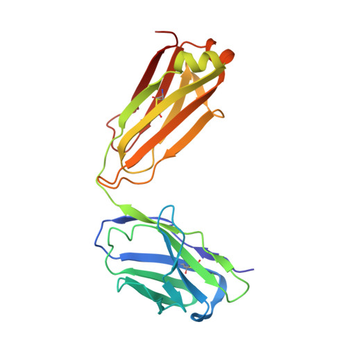
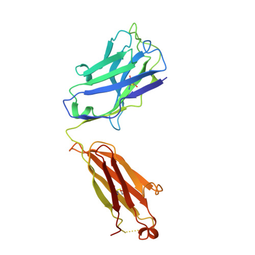
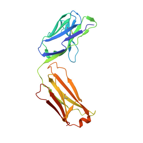
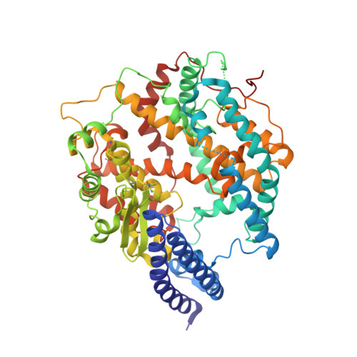
    7L0N

  • PubMed Abstract: 

    SARS-CoV-2 can mutate and evade immunity, with consequences for efficacy of emerging vaccines and antibody therapeutics. Here, we demonstrate that the immunodominant SARS-CoV-2 spike (S) receptor binding motif (RBM) is a highly variable region of S and provide epidemiological, clinical, and molecular characterization of a prevalent, sentinel RBM mutation, N439K. We demonstrate N439K S protein has enhanced binding affinity to the hACE2 receptor, and N439K viruses have similar in vitro replication fitness and cause infections with similar clinical outcomes as compared to wild type. We show the N439K mutation confers resistance against several neutralizing monoclonal antibodies, including one authorized for emergency use by the US Food and Drug Administration (FDA), and reduces the activity of some polyclonal sera from persons recovered from infection. Immune evasion mutations that maintain virulence and fitness such as N439K can emerge within SARS-CoV-2 S, highlighting the need for ongoing molecular surveillance to guide development and usage of vaccines and therapeutics.


  • Organizational Affiliation
    • MRC-University of Glasgow Centre for Virus Research, University of Glasgow, Glasgow G61 1QH, UK; Department of Clinical Research, London School of Hygiene and Tropical Medicine, London WC1E 7HT, UK.

Macromolecule Content 

  • Total Structure Weight: 380.02 kDa 
  • Atom Count: 26,468 
  • Modeled Residue Count: 3,304 
  • Deposited Residue Count: 3,366 
  • Unique protein chains: 6

Macromolecules

Find similar proteins by:|  3D Structure
Entity ID: 1
MoleculeChains  Sequence LengthOrganismDetailsImage
Monoclonal antibody S309 Fab light chainA [auth B],
C [auth D]
214Homo sapiensMutation(s): 0 
Find similar proteins by:|  3D Structure
Entity ID: 2
MoleculeChains  Sequence LengthOrganismDetailsImage
Monoclonal antibody S309 Fab heavy chainB [auth A],
D [auth C]
230Homo sapiensMutation(s): 0 
Find similar proteins by:|  3D Structure
Entity ID: 3
MoleculeChains  Sequence LengthOrganismDetailsImage
Monoclonal antibody S304 Fab light chainE [auth L],
G [auth N]
215Homo sapiensMutation(s): 0 
Find similar proteins by:|  3D Structure
Entity ID: 4
MoleculeChains  Sequence LengthOrganismDetailsImage
Monoclonal antibody S304 Fab heavy chainF [auth H],
H [auth M]
223Homo sapiensMutation(s): 0 
Find similar proteins by:|  3D Structure
Entity ID: 5
MoleculeChains  Sequence LengthOrganismDetailsImage
Angiotensin-converting enzyme 2I [auth E],
J [auth F]
597Homo sapiensMutation(s): 0 
Gene Names: ACE2UNQ868/PRO1885
EC: 3.4.17.23 (PDB Primary Data), 3.4.17 (UniProt)
UniProt & NIH Common Fund Data Resources
Find proteins for Q9BYF1 (Homo sapiens)
Explore Q9BYF1 
Go to UniProtKB:  Q9BYF1
PHAROS:  Q9BYF1
GTEx:  ENSG00000130234 
Entity Groups
Sequence Clusters30% Identity50% Identity70% Identity90% Identity95% Identity100% Identity
UniProt GroupQ9BYF1
Glycosylation
Glycosylation Sites: 5Go to GlyGen: Q9BYF1-1
Sequence Annotations
Expand
Reference Sequence
Find similar proteins by:|  3D Structure
Entity ID: 6
MoleculeChains  Sequence LengthOrganismDetailsImage
Spike protein S1K [auth R],
L [auth S]
204Severe acute respiratory syndrome coronavirus 2Mutation(s): 1 
Gene Names: S2
UniProt
Find proteins for P0DTC2 (Severe acute respiratory syndrome coronavirus 2)
Explore P0DTC2 
Go to UniProtKB:  P0DTC2
Entity Groups
Sequence Clusters30% Identity50% Identity70% Identity90% Identity95% Identity100% Identity
UniProt GroupP0DTC2
Glycosylation
Glycosylation Sites: 1Go to GlyGen: P0DTC2-1
Sequence Annotations
Expand
Reference Sequence

Oligosaccharides

Help  
Entity ID: 7
MoleculeChains Length2D Diagram GlycosylationD Interactions
alpha-D-mannopyranose-(1-6)-alpha-D-mannopyranose-(1-6)-beta-D-mannopyranose-(1-4)-2-acetamido-2-deoxy-beta-D-glucopyranose-(1-4)-[alpha-L-fucopyranose-(1-6)]2-acetamido-2-deoxy-beta-D-glucopyranoseM [auth G]6N-Glycosylation
Glycosylation Resources
GlyTouCan: G52146XU
GlyCosmos: G52146XU
GlyGen: G52146XU
Entity ID: 8
MoleculeChains Length2D Diagram GlycosylationD Interactions
alpha-D-mannopyranose-(1-6)-beta-D-mannopyranose-(1-4)-2-acetamido-2-deoxy-beta-D-glucopyranose-(1-4)-[alpha-L-fucopyranose-(1-6)]2-acetamido-2-deoxy-beta-D-glucopyranoseN [auth I]5N-Glycosylation
Glycosylation Resources
GlyTouCan: G00395TQ
GlyCosmos: G00395TQ
GlyGen: G00395TQ

Small Molecules

Ligands 8 Unique
IDChains Name / Formula / InChI Key2D Diagram3D Interactions
NAG

Query on NAG


Download:Ideal Coordinates CCD File
LA [auth E]
MA [auth E]
NA [auth E]
OA [auth E]
SA [auth F]
LA [auth E],
MA [auth E],
NA [auth E],
OA [auth E],
SA [auth F],
TA [auth F],
UA [auth F],
VA [auth F],
WA [auth F]
2-acetamido-2-deoxy-beta-D-glucopyranose
C8 H15 N O6
OVRNDRQMDRJTHS-FMDGEEDCSA-N
PG4

Query on PG4


Download:Ideal Coordinates CCD File
EA [auth N]TETRAETHYLENE GLYCOL
C8 H18 O5
UWHCKJMYHZGTIT-UHFFFAOYSA-N
PG5

Query on PG5


Download:Ideal Coordinates CCD File
V [auth H]1-METHOXY-2-[2-(2-METHOXY-ETHOXY]-ETHANE
C8 H18 O4
YFNKIDBQEZZDLK-UHFFFAOYSA-N
PGE

Query on PGE


Download:Ideal Coordinates CCD File
JB [auth S],
PA [auth E]
TRIETHYLENE GLYCOL
C6 H14 O4
ZIBGPFATKBEMQZ-UHFFFAOYSA-N
SO4

Query on SO4


Download:Ideal Coordinates CCD File
BA [auth N],
T [auth L]
SULFATE ION
O4 S
QAOWNCQODCNURD-UHFFFAOYSA-L
ZN

Query on ZN


Download:Ideal Coordinates CCD File
KA [auth E],
RA [auth F]
ZINC ION
Zn
PTFCDOFLOPIGGS-UHFFFAOYSA-N
CL

Query on CL


Download:Ideal Coordinates CCD File
AA [auth H]
AB [auth F]
CA [auth N]
CB [auth F]
DA [auth N]
AA [auth H],
AB [auth F],
CA [auth N],
CB [auth F],
DA [auth N],
DB [auth F],
EB [auth F],
FA [auth M],
FB [auth R],
GA [auth M],
GB [auth R],
HA [auth M],
IA [auth M],
IB [auth S],
JA [auth M],
KB [auth S],
LB [auth S],
O [auth B],
P [auth A],
Q [auth D],
QA [auth E],
R [auth C],
S [auth C],
W [auth H],
XA [auth F],
Z [auth H],
ZA [auth F]
CHLORIDE ION
Cl
VEXZGXHMUGYJMC-UHFFFAOYSA-M
NA

Query on NA


Download:Ideal Coordinates CCD File
BB [auth F]
HB [auth R]
U [auth H]
X [auth H]
Y [auth H]
BB [auth F],
HB [auth R],
U [auth H],
X [auth H],
Y [auth H],
YA [auth F]
SODIUM ION
Na
FKNQFGJONOIPTF-UHFFFAOYSA-N

Experimental Data & Validation

Experimental Data

  • Method: X-RAY DIFFRACTION
  • Resolution: 2.78 Å
  • R-Value Free:  0.325 (Depositor), 0.326 (DCC) 
  • R-Value Work:  0.290 (Depositor), 0.291 (DCC) 
  • R-Value Observed: 0.292 (Depositor) 
Space Group: P 1 21 1
Unit Cell:
Length ( Å )Angle ( ˚ )
a = 77.98α = 90
b = 186.58β = 96.4
c = 194.52γ = 90
Software Package:
Software NamePurpose
REFMACrefinement
Aimlessdata scaling
PHASERphasing
PDB_EXTRACTdata extraction
XDSdata reduction

Structure Validation

View Full Validation Report


Ligand Structure Quality Assessment 


Entry History 

Deposition Data

Revision History  (Full details and data files)

  • Version 1.0: 2021-02-17
    Type: Initial release
  • Version 1.1: 2021-03-10
    Changes: Database references
  • Version 1.2: 2021-03-17
    Changes: Database references
  • Version 1.3: 2023-10-18
    Changes: Data collection, Database references, Refinement description
  • Version 1.4: 2024-10-23
    Changes: Structure summary