7KX4 | pdb_00007kx4

Anti-CCHFV ADI-36121 Fab

  • Classification: IMMUNE SYSTEM
  • Organism(s): Homo sapiens
  • Expression System: Homo sapiens
  • Mutation(s): No 

  • Deposited: 2020-12-03 Released: 2021-12-01 
  • Deposition Author(s): Mishra, A.K., McLellan, J.S.
  • Funding Organization(s): National Institutes of Health/National Institute Of Allergy and Infectious Diseases (NIH/NIAID)

Experimental Data Snapshot

  • Method: X-RAY DIFFRACTION
  • Resolution: 2.60 Å
  • R-Value Free: 
    0.239 (Depositor), 0.238 (DCC) 
  • R-Value Work: 
    0.198 (Depositor), 0.198 (DCC) 
  • R-Value Observed: 
    0.200 (Depositor) 

Starting Models: experimental
View more details

wwPDB Validation 3D Report Full Report

Validation slider image for 7KX4

This is version 1.3 of the entry. See complete history

Literature

Structural basis of synergistic neutralization of Crimean-Congo hemorrhagic fever virus by human antibodies.

Mishra, A.K.Hellert, J.Freitas, N.Guardado-Calvo, P.Haouz, A.Fels, J.M.Maurer, D.P.Abelson, D.M.Bornholdt, Z.A.Walker, L.M.Chandran, K.Cosset, F.L.McLellan, J.S.Rey, F.A.

(2022) Science 375: 104-109

  • DOI: https://doi.org/10.1126/science.abl6502
  • Primary Citation Related Structures: 
    7A59, 7A5A, 7KX4, 7L7R

  • PubMed Abstract: 

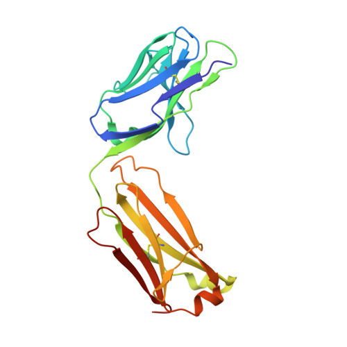
    Crimean-Congo hemorrhagic fever virus (CCHFV) is the most widespread tick-borne zoonotic virus, with a 30% case fatality rate in humans. Structural information is lacking in regard to the CCHFV membrane fusion glycoprotein Gc—the main target of the host neutralizing antibody response—as well as antibody–mediated neutralization mechanisms. We describe the structure of prefusion Gc bound to the antigen-binding fragments (Fabs) of two neutralizing antibodies that display synergy when combined, as well as the structure of trimeric, postfusion Gc. The structures show the two Fabs acting in concert to block membrane fusion, with one targeting the fusion loops and the other blocking Gc trimer formation. The structures also revealed the neutralization mechanism of previously reported antibodies against CCHFV, providing the molecular underpinnings essential for developing CCHFV–specific medical countermeasures for epidemic preparedness.


  • Organizational Affiliation
    • Department of Molecular Biosciences, The University of Texas at Austin, Austin, TX 78712, USA.

Macromolecule Content 

  • Total Structure Weight: 96.16 kDa 
  • Atom Count: 6,842 
  • Modeled Residue Count: 856 
  • Deposited Residue Count: 888 
  • Unique protein chains: 2

Macromolecules

Find similar proteins by:|  3D Structure
Entity ID: 1
MoleculeChains  Sequence LengthOrganismDetailsImage
ADI-36121 Fab light chain
A, C
214Homo sapiensMutation(s): 0 
Find similar proteins by:|  3D Structure
Entity ID: 2
MoleculeChains  Sequence LengthOrganismDetailsImage
ADI-36121 Fab heavy chain
B, D
230Homo sapiensMutation(s): 0 

Experimental Data & Validation

Experimental Data

  • Method: X-RAY DIFFRACTION
  • Resolution: 2.60 Å
  • R-Value Free:  0.239 (Depositor), 0.238 (DCC) 
  • R-Value Work:  0.198 (Depositor), 0.198 (DCC) 
  • R-Value Observed: 0.200 (Depositor) 
Space Group: P 21 21 21
Unit Cell:
Length ( Å )Angle ( ˚ )
a = 58.19α = 90
b = 76.64β = 90
c = 208.52γ = 90
Software Package:
Software NamePurpose
PHENIXrefinement
MOSFLMdata reduction
Aimlessdata scaling
PHASERphasing
PDB_EXTRACTdata extraction

Structure Validation

View Full Validation Report


Entry History 

& Funding Information

Deposition Data


Funding OrganizationLocationGrant Number
National Institutes of Health/National Institute Of Allergy and Infectious Diseases (NIH/NIAID)United StatesU19 AI142777

Revision History  (Full details and data files)

  • Version 1.0: 2021-12-01
    Type: Initial release
  • Version 1.1: 2022-01-19
    Changes: Database references
  • Version 1.2: 2023-10-18
    Changes: Data collection, Refinement description
  • Version 1.3: 2024-10-23
    Changes: Structure summary