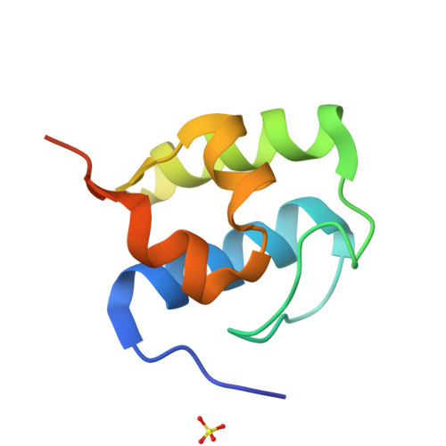
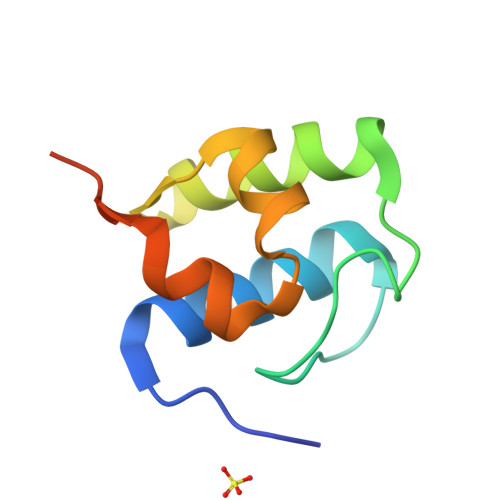
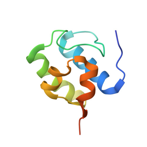
7KW3

Non Ribosomal PCP domain


Experimental Data Snapshot

  • Method: X-RAY DIFFRACTION
  • Resolution: 2.30 Å
  • R-Value Free: 0.206 
  • R-Value Work: 0.184 
  • R-Value Observed: 0.185 

Starting Model: in silico
View more details

wwPDB Validation   3D Report Full Report


This is version 2.3 of the entry. See complete history


Literature

Structures of a non-ribosomal peptide synthetase condensation domain suggest the basis of substrate selectivity.

Izore, T.Candace Ho, Y.T.Kaczmarski, J.A.Gavriilidou, A.Chow, K.H.Steer, D.L.Goode, R.J.A.Schittenhelm, R.B.Tailhades, J.Tosin, M.Challis, G.L.Krenske, E.H.Ziemert, N.Jackson, C.J.Cryle, M.J.

(2021) Nat Commun 12: 2511-2511

  • DOI: https://doi.org/10.1038/s41467-021-22623-0
  • Primary Citation of Related Structures:  
    7KVW, 7KW0, 7KW2, 7KW3

  • PubMed Abstract: 

    Non-ribosomal peptide synthetases are important enzymes for the assembly of complex peptide natural products. Within these multi-modular assembly lines, condensation domains perform the central function of chain assembly, typically by forming a peptide bond between two peptidyl carrier protein (PCP)-bound substrates. In this work, we report structural snapshots of a condensation domain in complex with an aminoacyl-PCP acceptor substrate. These structures allow the identification of a mechanism that controls access of acceptor substrates to the active site in condensation domains. The structures of this complex also allow us to demonstrate that condensation domain active sites do not contain a distinct pocket to select the side chain of the acceptor substrate during peptide assembly but that residues within the active site motif can instead serve to tune the selectivity of these central biosynthetic domains.


  • Organizational Affiliation

    Department of Biochemistry and Molecular Biology, The Monash Biomedicine Discovery Institute, Monash University, Clayton, VIC, Australia. thierry.izore@monash.edu.


Macromolecules
Find similar proteins by:  (by identity cutoff)  |  3D Structure
Entity ID: 1
MoleculeChains Sequence LengthOrganismDetailsImage
PCP domain87Thermobifida fusca YXMutation(s): 0 
Gene Names: Tfu_1867
UniProt
Find proteins for Q47NR9 (Thermobifida fusca (strain YX))
Explore Q47NR9 
Go to UniProtKB:  Q47NR9
Entity Groups  
Sequence Clusters30% Identity50% Identity70% Identity90% Identity95% Identity100% Identity
UniProt GroupQ47NR9
Sequence Annotations
Expand
  • Reference Sequence
Small Molecules
Ligands 1 Unique
IDChains Name / Formula / InChI Key2D Diagram3D Interactions
SO4
Query on SO4

Download Ideal Coordinates CCD File 
B [auth A]SULFATE ION
O4 S
QAOWNCQODCNURD-UHFFFAOYSA-L
Experimental Data & Validation

Experimental Data

  • Method: X-RAY DIFFRACTION
  • Resolution: 2.30 Å
  • R-Value Free: 0.206 
  • R-Value Work: 0.184 
  • R-Value Observed: 0.185 
  • Space Group: P 43 3 2
Unit Cell:
Length ( Å )Angle ( ˚ )
a = 100.914α = 90
b = 100.914β = 90
c = 100.914γ = 90
Software Package:
Software NamePurpose
PHENIXrefinement
XDSdata reduction
Aimlessdata scaling
PHENIXphasing

Structure Validation

View Full Validation Report


Entry History & Funding Information

Deposition Data


Funding OrganizationLocationGrant Number
Australian Research Council (ARC)AustraliaDP190101272
National Health and Medical Research Council (NHMRC, Australia)AustraliaAPP1140619

Revision History  (Full details and data files)

  • Version 1.0: 2021-03-24
    Type: Initial release
  • Version 2.0: 2021-04-21
    Changes: Advisory, Atomic model, Data collection, Database references, Derived calculations, Experimental preparation, Non-polymer description, Polymer sequence, Source and taxonomy, Structure summary
  • Version 2.1: 2021-05-19
    Changes: Database references
  • Version 2.2: 2024-03-06
    Changes: Data collection, Database references
  • Version 2.3: 2024-04-03
    Changes: Refinement description