7KVA | pdb_00007kva

Structure of West Nile virus (Kunjin)


Experimental Data Snapshot

  • Method: ELECTRON MICROSCOPY
  • Resolution: 3.10 Å
  • Aggregation State: PARTICLE 
  • Reconstruction Method: SINGLE PARTICLE 

Starting Model: experimental
View more details

wwPDB Validation 3D Report Full Report

Validation slider image for 7KVA

This is version 1.2 of the entry. See complete history

Literature

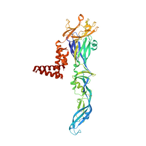
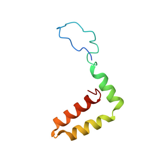
A unified route for flavivirus structures uncovers essential pocket factors conserved across pathogenic viruses.

Hardy, J.M.Newton, N.D.Modhiran, N.Scott, C.A.P.Venugopal, H.Vet, L.J.Young, P.R.Hall, R.A.Hobson-Peters, J.Coulibaly, F.Watterson, D.

(2021) Nat Commun 12: 3266-3266

  • DOI: https://doi.org/10.1038/s41467-021-22773-1
  • Primary Citation Related Structures: 
    7KV8, 7KV9, 7KVA, 7KVB

  • PubMed Abstract: 

    The epidemic emergence of relatively rare and geographically isolated flaviviruses adds to the ongoing disease burden of viruses such as dengue. Structural analysis is key to understand and combat these pathogens. Here, we present a chimeric platform based on an insect-specific flavivirus for the safe and rapid structural analysis of pathogenic viruses. We use this approach to resolve the architecture of two neurotropic viruses and a structure of dengue virus at 2.5  Å, the highest resolution for an enveloped virion. These reconstructions allow improved modelling of the stem region of the envelope protein, revealing two lipid-like ligands within highly conserved pockets. We show that these sites are essential for viral growth and important for viral maturation. These findings define a hallmark of flavivirus virions and a potential target for broad-spectrum antivirals and vaccine design. We anticipate the chimeric platform to be widely applicable for investigating flavivirus biology.


  • Organizational Affiliation
    • Infection and Immunity Program, Biomedicine Discovery Institute and Department of Biochemistry and Molecular Biology, Monash University, Clayton, VIC, Australia.

Macromolecule Content 

  • Total Structure Weight: 186.78 kDa 
  • Atom Count: 12,877 
  • Modeled Residue Count: 1,716 
  • Deposited Residue Count: 1,728 
  • Unique protein chains: 2

Macromolecules

Find similar proteins by:|  3D Structure
Entity ID: 1
MoleculeChains  Sequence LengthOrganismDetailsImage
Envelope protein E
A, B, C
501Kunjin virusMutation(s): 0 
UniProt
Find proteins for P14335 (Kunjin virus (strain MRM61C))
Explore P14335 
Go to UniProtKB:  P14335
Entity Groups
Sequence Clusters30% Identity50% Identity70% Identity90% Identity95% Identity100% Identity
UniProt GroupP14335
Glycosylation
Glycosylation Sites: 1
Sequence Annotations
Expand
Reference Sequence
Find similar proteins by:|  3D Structure
Entity ID: 2
MoleculeChains  Sequence LengthOrganismDetailsImage
Matrix protein MD [auth a],
E [auth b],
F [auth c]
75Kunjin virusMutation(s): 0 
UniProt
Find proteins for P14335 (Kunjin virus (strain MRM61C))
Explore P14335 
Go to UniProtKB:  P14335
Entity Groups
Sequence Clusters30% Identity50% Identity70% Identity90% Identity95% Identity100% Identity
UniProt GroupP14335
Sequence Annotations
Expand
Reference Sequence

Experimental Data & Validation

Experimental Data

  • Method: ELECTRON MICROSCOPY
  • Resolution: 3.10 Å
  • Aggregation State: PARTICLE 
  • Reconstruction Method: SINGLE PARTICLE 
EM Software:
TaskSoftware PackageVersion
MODEL REFINEMENTPHENIX1.16
RECONSTRUCTIONRELION3.1

Structure Validation

View Full Validation Report


Entry History 

& Funding Information

Deposition Data


Funding OrganizationLocationGrant Number
National Health and Medical Research Council (NHMRC, Australia)AustraliaAPP1164216

Revision History  (Full details and data files)

  • Version 1.0: 2020-12-23
    Type: Initial release
  • Version 1.1: 2021-07-14
    Changes: Database references
  • Version 1.2: 2024-10-30
    Changes: Data collection, Database references, Derived calculations, Refinement description, Structure summary