7KV7 | pdb_00007kv7

Surface glycan-binding protein B from Bacteroides fluxus in complex with laminaritriose


Experimental Data Snapshot

  • Method: X-RAY DIFFRACTION
  • Resolution: 1.76 Å
  • R-Value Free: 
    0.202 (Depositor), 0.202 (DCC) 
  • R-Value Work: 
    0.159 (Depositor), 0.159 (DCC) 

wwPDB Validation 3D Report Full Report

Validation slider image for 7KV7

Ligand Structure Quality Assessment 


This is version 1.3 of the entry. See complete history

Literature

Distinct protein architectures mediate species-specific beta-glucan binding and metabolism in the human gut microbiota.

Tamura, K.Dejean, G.Van Petegem, F.Brumer, H.

(2021) J Biological Chem 296: 100415-100415

  • DOI: https://doi.org/10.1016/j.jbc.2021.100415
  • Primary Citation Related Structures: 
    7KV1, 7KV2, 7KV3, 7KV4, 7KV5, 7KV6, 7KV7, 7KWB, 7KWC

  • PubMed Abstract: 

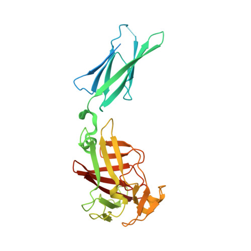
    Complex glycans that evade our digestive system are major nutrients that feed the human gut microbiota (HGM). The prevalence of Bacteroidetes in the HGM of populations worldwide is engendered by the evolution of polysaccharide utilization loci (PULs), which encode concerted protein systems to utilize the myriad complex glycans in our diets. Despite their crucial roles in glycan recognition and transport, cell-surface glycan-binding proteins (SGBPs) remained understudied cogs in the PUL machinery. Here, we report the structural and biochemical characterization of a suite of SGBP-A and SGBP-B structures from three syntenic β(1,3)-glucan utilization loci (1,3GULs) from Bacteroides thetaiotaomicron (Bt), Bacteroides uniformis (Bu), and B. fluxus (Bf), which have varying specificities for distinct β-glucans. Ligand complexes provide definitive insight into β(1,3)-glucan selectivity in the HGM, including structural features enabling dual β(1,3)-glucan/mixed-linkage β(1,3)/β(1,4)-glucan-binding capability in some orthologs. The tertiary structural conservation of SusD-like SGBPs-A is juxtaposed with the diverse architectures and binding modes of the SGBPs-B. Specifically, the structures of the trimodular BtSGBP-B and BuSGBP-B revealed a tandem repeat of carbohydrate-binding module-like domains connected by long linkers. In contrast, BfSGBP-B comprises a bimodular architecture with a distinct β-barrel domain at the C terminus that bears a shallow binding canyon. The molecular insights obtained here contribute to our fundamental understanding of HGM function, which in turn may inform tailored microbial intervention therapies.


  • Organizational Affiliation
    • Michael Smith Laboratories, University of British Columbia, Vancouver, British Columbia, Canada; Department of Biochemistry and Molecular Biology, The Life Sciences Institute, University of British Columbia, Vancouver, British Columbia, Canada.

Macromolecule Content 

  • Total Structure Weight: 63.76 kDa 
  • Atom Count: 4,590 
  • Modeled Residue Count: 506 
  • Deposited Residue Count: 580 
  • Unique protein chains: 1

Macromolecules

Find similar proteins by:|  3D Structure
Entity ID: 1
MoleculeChains  Sequence LengthOrganismDetailsImage
PKD domain proteinA [auth AAA],
B [auth BBB]
290Bacteroides fluxus YIT 12057Mutation(s): 0 
Gene Names: HMPREF9446_00613
UniProt
Find proteins for F3PPH3 (Bacteroides fluxus YIT 12057)
Explore F3PPH3 
Go to UniProtKB:  F3PPH3
Entity Groups
Sequence Clusters30% Identity50% Identity70% Identity90% Identity95% Identity100% Identity
UniProt GroupF3PPH3
Sequence Annotations
Expand
Reference Sequence

Oligosaccharides

Help  
Entity ID: 2
MoleculeChains Length2D Diagram GlycosylationD Interactions
beta-D-glucopyranose-(1-3)-beta-D-glucopyranoseC [auth BbB]2N/A
Glycosylation Resources
GlyTouCan: G36535HU
GlyCosmos: G36535HU

Small Molecules

Ligands 5 Unique
IDChains Name / Formula / InChI Key2D Diagram3D Interactions
BGC
(Subject of Investigation/LOI)

Query on BGC


Download:Ideal Coordinates CCD File
E [auth AAA],
F [auth AAA],
I [auth BBB]
beta-D-glucopyranose
C6 H12 O6
WQZGKKKJIJFFOK-VFUOTHLCSA-N
LYS

Query on LYS


Download:Ideal Coordinates CCD File
D [auth AAA]LYSINE
C6 H15 N2 O2
KDXKERNSBIXSRK-YFKPBYRVSA-O
PEG

Query on PEG


Download:Ideal Coordinates CCD File
G [auth AAA]DI(HYDROXYETHYL)ETHER
C4 H10 O3
MTHSVFCYNBDYFN-UHFFFAOYSA-N
SO4

Query on SO4


Download:Ideal Coordinates CCD File
H [auth AAA],
K [auth BBB]
SULFATE ION
O4 S
QAOWNCQODCNURD-UHFFFAOYSA-L
EDO

Query on EDO


Download:Ideal Coordinates CCD File
J [auth BBB]1,2-ETHANEDIOL
C2 H6 O2
LYCAIKOWRPUZTN-UHFFFAOYSA-N

Biologically Interesting Molecules (External Reference) 

1 Unique

Experimental Data & Validation

Experimental Data

  • Method: X-RAY DIFFRACTION
  • Resolution: 1.76 Å
  • R-Value Free:  0.202 (Depositor), 0.202 (DCC) 
  • R-Value Work:  0.159 (Depositor), 0.159 (DCC) 
Space Group: I 1 2 1
Unit Cell:
Length ( Å )Angle ( ˚ )
a = 74.268α = 90
b = 59.249β = 102.611
c = 148.137γ = 90
Software Package:
Software NamePurpose
REFMACrefinement
XDSdata reduction
Aimlessdata scaling
PHASERphasing

Structure Validation

View Full Validation Report


Ligand Structure Quality Assessment 


Entry History 

& Funding Information

Deposition Data


Funding OrganizationLocationGrant Number
Canadian Institutes of Health Research (CIHR)Canada--

Revision History  (Full details and data files)

  • Version 1.0: 2021-02-17
    Type: Initial release
  • Version 1.1: 2021-03-03
    Changes: Database references
  • Version 1.2: 2021-07-21
    Changes: Database references
  • Version 1.3: 2024-03-06
    Changes: Data collection, Database references