7KPA | pdb_00007kpa

asymmetric hTNF-alpha


Experimental Data Snapshot

  • Method: X-RAY DIFFRACTION
  • Resolution: 2.30 Å
  • R-Value Free: 
    0.262 (Depositor), 0.263 (DCC) 
  • R-Value Work: 
    0.216 (Depositor), 0.216 (DCC) 
  • R-Value Observed: 
    0.221 (Depositor) 

Starting Model: other
View more details

wwPDB Validation 3D Report Full Report

Validation slider image for 7KPA

Ligand Structure Quality Assessment 


This is version 1.4 of the entry. See complete history

Literature

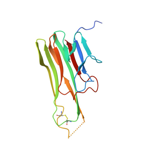
A conformation-selective monoclonal antibody against a small molecule-stabilised signalling-deficient form of TNF.

Lightwood, D.J.Munro, R.J.Porter, J.McMillan, D.Carrington, B.Turner, A.Scott-Tucker, A.Hickford, E.S.Schmidt, A.Fox III, D.Maloney, A.Ceska, T.Bourne, T.O'Connell, J.Lawson, A.D.G.

(2021) Nat Commun 12: 583-583

  • DOI: https://doi.org/10.1038/s41467-020-20825-6
  • Primary Citation Related Structures: 
    7KPA, 7KPB

  • PubMed Abstract: 

    We have recently described the development of a series of small-molecule inhibitors of human tumour necrosis factor (TNF) that stabilise an open, asymmetric, signalling-deficient form of the soluble TNF trimer. Here, we describe the generation, characterisation, and utility of a monoclonal antibody that selectively binds with high affinity to the asymmetric TNF trimer-small molecule complex. The antibody helps to define the molecular dynamics of the apo TNF trimer, reveals the mode of action and specificity of the small molecule inhibitors, acts as a chaperone in solving the human TNF-TNFR1 complex crystal structure, and facilitates the measurement of small molecule target occupancy in complex biological samples. We believe this work defines a role for monoclonal antibodies as tools to facilitate the discovery and development of small-molecule inhibitors of protein-protein interactions.


  • Organizational Affiliation
    • UCB Pharma, 208 Bath Road, Slough, SL1 3WE, UK. Daniel.Lightwood@ucb.com.

Macromolecule Content 

  • Total Structure Weight: 52.93 kDa 
  • Atom Count: 3,275 
  • Modeled Residue Count: 426 
  • Deposited Residue Count: 474 
  • Unique protein chains: 1

Macromolecules

Find similar proteins by:|  3D Structure
Entity ID: 1
MoleculeChains  Sequence LengthOrganismDetailsImage
Tumor necrosis factor
A, B, C
158Homo sapiensMutation(s): 0 
Gene Names: TNFTNFATNFSF2
UniProt & NIH Common Fund Data Resources
Find proteins for P01375 (Homo sapiens)
Explore P01375 
Go to UniProtKB:  P01375
PHAROS:  P01375
GTEx:  ENSG00000232810 
Entity Groups
Sequence Clusters30% Identity50% Identity70% Identity90% Identity95% Identity100% Identity
UniProt GroupP01375
Sequence Annotations
Expand
Reference Sequence

Small Molecules

Ligands 1 Unique
IDChains Name / Formula / InChI Key2D Diagram3D Interactions
D84
(Subject of Investigation/LOI)

Query on D84


Download:Ideal Coordinates CCD File
D [auth C]5-(1-{[2-(difluoromethoxy)phenyl]methyl}-2-{[3-(2-oxopyrrolidin-1-yl)phenoxy]methyl}-1H-benzimidazol-6-yl)pyridin-2(1H)-one
C31 H26 F2 N4 O4
DNTKSNFCNRDJAC-UHFFFAOYSA-N

Experimental Data & Validation

Experimental Data

  • Method: X-RAY DIFFRACTION
  • Resolution: 2.30 Å
  • R-Value Free:  0.262 (Depositor), 0.263 (DCC) 
  • R-Value Work:  0.216 (Depositor), 0.216 (DCC) 
  • R-Value Observed: 0.221 (Depositor) 
Space Group: P 21 21 21
Unit Cell:
Length ( Å )Angle ( ˚ )
a = 55.13α = 90
b = 82.13β = 90
c = 93.04γ = 90
Software Package:
Software NamePurpose
PHENIXrefinement
PDB_EXTRACTdata extraction
XDSdata reduction
XSCALEdata scaling
REFMACphasing

Structure Validation

View Full Validation Report


Ligand Structure Quality Assessment 


Entry History 

Deposition Data

Revision History  (Full details and data files)

  • Version 1.0: 2021-01-13
    Type: Initial release
  • Version 1.1: 2021-02-03
    Changes: Database references
  • Version 1.2: 2022-06-29
    Changes: Database references, Structure summary
  • Version 1.3: 2024-04-03
    Changes: Data collection, Refinement description
  • Version 1.4: 2024-11-06
    Changes: Structure summary