7KOX | pdb_00007kox

Alpha-7 nicotinic acetylcholine receptor bound to epibatidine and PNU-120596 in the activated state


Experimental Data Snapshot

  • Method: ELECTRON MICROSCOPY
  • Resolution: 2.70 Å
  • Aggregation State: PARTICLE 
  • Reconstruction Method: SINGLE PARTICLE 

wwPDB Validation 3D Report Full Report

Validation slider image for 7KOX

This is version 2.3 of the entry. See complete history

Literature

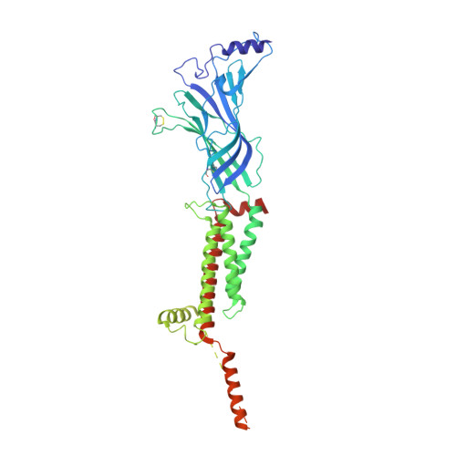
Structure and gating mechanism of the alpha 7 nicotinic acetylcholine receptor.

Noviello, C.M.Gharpure, A.Mukhtasimova, N.Cabuco, R.Baxter, L.Borek, D.Sine, S.M.Hibbs, R.E.

(2021) Cell 184: 2121

  • DOI: https://doi.org/10.1016/j.cell.2021.02.049
  • Primary Citation Related Structures: 
    7KOO, 7KOQ, 7KOX

  • PubMed Abstract: 

    The α7 nicotinic acetylcholine receptor plays critical roles in the central nervous system and in the cholinergic inflammatory pathway. This ligand-gated ion channel assembles as a homopentamer, is exceptionally permeable to Ca 2+ , and desensitizes faster than any other Cys-loop receptor. The α7 receptor has served as a prototype for the Cys-loop superfamily yet has proven refractory to structural analysis. We present cryo-EM structures of the human α7 nicotinic receptor in a lipidic environment in resting, activated, and desensitized states, illuminating the principal steps in the gating cycle. The structures also reveal elements that contribute to its function, including a C-terminal latch that is permissive for channel opening, and an anionic ring in the extracellular vestibule that contributes to its high conductance and calcium permeability. Comparisons among the α7 structures provide a foundation for mapping the gating cycle and reveal divergence in gating mechanisms in the Cys-loop receptor superfamily.


  • Organizational Affiliation
    • Department of Neuroscience, Peter O'Donnell Jr. Brain Institute, University of Texas Southwestern Medical Center, Dallas, TX 75390, USA; Department of Biophysics, University of Texas Southwestern Medical Center, Dallas, TX 75390, USA.

Macromolecule Content 

  • Total Structure Weight: 324.74 kDa 
  • Atom Count: 15,940 
  • Modeled Residue Count: 1,930 
  • Deposited Residue Count: 2,810 
  • Unique protein chains: 1

Macromolecules

Find similar proteins by:|  3D Structure
Entity ID: 1
MoleculeChains  Sequence LengthOrganismDetailsImage
Neuronal acetylcholine receptor subunit alpha-7,Soluble cytochrome b562 fusion
A, B, C, D, E
562Homo sapiensEscherichia coliMutation(s): 3 
Gene Names: CHRNA7NACHRA7cybC
Membrane Entity: Yes 
UniProt & NIH Common Fund Data Resources
Find proteins for P0ABE7 (Escherichia coli)
Explore P0ABE7 
Go to UniProtKB:  P0ABE7
Find proteins for P36544 (Homo sapiens)
Explore P36544 
Go to UniProtKB:  P36544
PHAROS:  P36544
GTEx:  ENSG00000175344 
Entity Groups
Sequence Clusters30% Identity50% Identity70% Identity90% Identity95% Identity100% Identity
UniProt GroupsP36544P0ABE7
Glycosylation
Glycosylation Sites: 3Go to GlyGen: P36544-1
Sequence Annotations
Expand
Reference Sequence

Oligosaccharides

Help  
Entity ID: 2
MoleculeChains Length2D Diagram GlycosylationD Interactions
2-acetamido-2-deoxy-beta-D-glucopyranose-(1-4)-2-acetamido-2-deoxy-beta-D-glucopyranose
F, G, H, I, J
2N-Glycosylation
Glycosylation Resources
GlyTouCan: G42666HT
GlyCosmos: G42666HT
GlyGen: G42666HT

Small Molecules

Ligands 3 Unique
IDChains Name / Formula / InChI Key2D Diagram3D Interactions
NAG

Query on NAG


Download:Ideal Coordinates CCD File
AA [auth E]
BA [auth E]
K [auth A]
L [auth A]
O [auth B]
AA [auth E],
BA [auth E],
K [auth A],
L [auth A],
O [auth B],
P [auth B],
S [auth C],
T [auth C],
W [auth D],
X [auth D]
2-acetamido-2-deoxy-beta-D-glucopyranose
C8 H15 N O6
OVRNDRQMDRJTHS-FMDGEEDCSA-N
EPJ

Query on EPJ


Download:Ideal Coordinates CCD File
CA [auth E],
M [auth A],
Q [auth B],
U [auth C],
Y [auth D]
EPIBATIDINE
C11 H13 Cl N2
NLPRAJRHRHZCQQ-IVZWLZJFSA-N
CA

Query on CA


Download:Ideal Coordinates CCD File
DA [auth E],
N [auth A],
R [auth B],
V [auth C],
Z [auth D]
CALCIUM ION
Ca
BHPQYMZQTOCNFJ-UHFFFAOYSA-N
Binding Affinity Annotations 
IDSourceBinding Affinity
EPJ BindingDB:  7KOX Ki: min: 2.2, max: 430 (nM) from 16 assay(s)
Kd: min: 1.06, max: 6.3 (nM) from 2 assay(s)
IC50: 31 (nM) from 1 assay(s)
EC50: min: 1300, max: 3500 (nM) from 2 assay(s)

Experimental Data & Validation

Experimental Data

  • Method: ELECTRON MICROSCOPY
  • Resolution: 2.70 Å
  • Aggregation State: PARTICLE 
  • Reconstruction Method: SINGLE PARTICLE 
EM Software:
TaskSoftware PackageVersion
MODEL REFINEMENTPHENIX

Structure Validation

View Full Validation Report


Entry History 

& Funding Information

Deposition Data


Funding OrganizationLocationGrant Number
National Institutes of Health/National Institute of Mental Health (NIH/NIMH)United StatesNS095899
National Institutes of Health/National Institute of Mental Health (NIH/NIMH)United StatesNS077983

Revision History  (Full details and data files)

  • Version 1.0: 2021-03-17
    Type: Initial release
  • Version 1.1: 2021-03-31
    Changes: Database references
  • Version 2.0: 2021-05-05
    Type: Coordinate replacement
    Reason: Sequence discrepancy
    Changes: Advisory, Atomic model, Data collection, Database references, Derived calculations, Polymer sequence, Refinement description, Structure summary
  • Version 2.1: 2021-05-12
    Changes: Database references
  • Version 2.2: 2024-10-23
    Changes: Data collection, Database references, Structure summary
  • Version 2.3: 2025-05-14
    Changes: Data collection