7KO8 | pdb_00007ko8

Cryo-EM structure of the mature and infective Mayaro virus


Experimental Data Snapshot

  • Method: ELECTRON MICROSCOPY
  • Resolution: 4.40 Å
  • Aggregation State: PARTICLE 
  • Reconstruction Method: SINGLE PARTICLE 

wwPDB Validation 3D Report Full Report

Validation slider image for 7KO8

This is version 1.2 of the entry. See complete history

Literature

Cryo-EM structure of the mature and infective Mayaro virus at 4.4 angstrom resolution reveals features of arthritogenic alphaviruses.

Ribeiro-Filho, H.V.Coimbra, L.D.Cassago, A.Rocha, R.P.F.Guerra, J.V.D.S.de Felicio, R.Carnieli, C.M.Leme, L.Padilha, A.C.Paes Leme, A.F.Trivella, D.B.B.Portugal, R.V.Lopes-de-Oliveira, P.S.Marques, R.E.

(2021) Nat Commun 12: 3038-3038

  • DOI: https://doi.org/10.1038/s41467-021-23400-9
  • Primary Citation Related Structures: 
    7KO8

  • PubMed Abstract: 

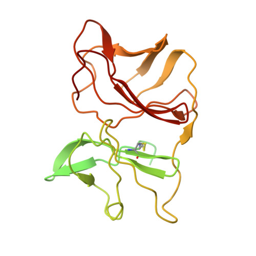
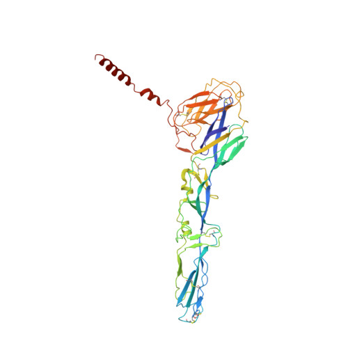
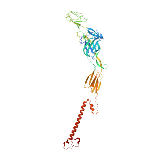
    Mayaro virus (MAYV) is an emerging arbovirus of the Americas that may cause a debilitating arthritogenic disease. The biology of MAYV is not fully understood and largely inferred from related arthritogenic alphaviruses. Here, we present the structure of MAYV at 4.4 Å resolution, obtained from a preparation of mature, infective virions. MAYV presents typical alphavirus features and organization. Interactions between viral proteins that lead to particle formation are described together with a hydrophobic pocket formed between E1 and E2 spike proteins and conformational epitopes specific of MAYV. We also describe MAYV glycosylation residues in E1 and E2 that may affect MXRA8 host receptor binding, and a molecular "handshake" between MAYV spikes formed by N262 glycosylation in adjacent E2 proteins. The structure of MAYV is suggestive of structural and functional complexity among alphaviruses, which may be targeted for specificity or antiviral activity.


  • Organizational Affiliation
    • Brazilian Biosciences National Laboratory, Brazilian Center for Research in Energy and Materials (CNPEM), Campinas, SP, Brazil.

Macromolecule Content 

  • Total Structure Weight: 499.37 kDa 
  • Atom Count: 31,153 
  • Modeled Residue Count: 4,004 
  • Deposited Residue Count: 4,464 
  • Unique protein chains: 3

Macromolecules

Find similar proteins by:|  3D Structure
Entity ID: 1
MoleculeChains  Sequence LengthOrganismDetailsImage
Capsid protein
A, D, G, J
258Mayaro virusMutation(s): 0 
UniProt
Find proteins for A0A0P0BSS5 (Mayaro virus)
Explore A0A0P0BSS5 
Go to UniProtKB:  A0A0P0BSS5
Entity Groups
Sequence Clusters30% Identity50% Identity70% Identity90% Identity95% Identity100% Identity
UniProt GroupA0A0P0BSS5
Sequence Annotations
Expand
Reference Sequence
Find similar proteins by:|  3D Structure
Entity ID: 2
MoleculeChains  Sequence LengthOrganismDetailsImage
E1 glycoproteinB,
E,
H,
K [auth L]
436Mayaro virusMutation(s): 0 
UniProt
Find proteins for A0A0P0CH64 (Mayaro virus)
Explore A0A0P0CH64 
Go to UniProtKB:  A0A0P0CH64
Entity Groups
Sequence Clusters30% Identity50% Identity70% Identity90% Identity95% Identity100% Identity
UniProt GroupA0A0P0CH64
Glycosylation
Glycosylation Sites: 1
Sequence Annotations
Expand
Reference Sequence
Find similar proteins by:|  3D Structure
Entity ID: 3
MoleculeChains  Sequence LengthOrganismDetailsImage
E2 glycoproteinC,
F,
I,
L [auth M]
422Mayaro virusMutation(s): 0 
UniProt
Find proteins for A0A2I4SMH8 (Mayaro virus)
Explore A0A2I4SMH8 
Go to UniProtKB:  A0A2I4SMH8
Entity Groups
Sequence Clusters30% Identity50% Identity70% Identity90% Identity95% Identity100% Identity
UniProt GroupA0A2I4SMH8
Glycosylation
Glycosylation Sites: 1
Sequence Annotations
Expand
Reference Sequence

Oligosaccharides

Help  
Entity ID: 4
MoleculeChains Length2D Diagram GlycosylationD Interactions
beta-D-mannopyranose-(1-4)-2-acetamido-2-deoxy-beta-D-glucopyranose-(1-4)-2-acetamido-2-deoxy-beta-D-glucopyranoseM [auth N],
O [auth P],
R [auth T]
3N-Glycosylation
Glycosylation Resources
GlyTouCan: G15407YE
GlyCosmos: G15407YE
GlyGen: G15407YE
Entity ID: 5
MoleculeChains Length2D Diagram GlycosylationD Interactions
alpha-D-mannopyranose-(1-3)-[alpha-D-mannopyranose-(1-6)]beta-D-mannopyranose-(1-4)-2-acetamido-2-deoxy-beta-D-glucopyranose-(1-4)-2-acetamido-2-deoxy-beta-D-glucopyranoseN [auth O],
P [auth Q],
S [auth U],
T [auth S]
5N-Glycosylation
Glycosylation Resources
GlyTouCan: G22768VO
GlyCosmos: G22768VO
GlyGen: G22768VO
Entity ID: 6
MoleculeChains Length2D Diagram GlycosylationD Interactions
2-acetamido-2-deoxy-beta-D-glucopyranose-(1-4)-2-acetamido-2-deoxy-beta-D-glucopyranoseQ [auth R]2N-Glycosylation
Glycosylation Resources
GlyTouCan: G42666HT
GlyCosmos: G42666HT
GlyGen: G42666HT

Experimental Data & Validation

Experimental Data

  • Method: ELECTRON MICROSCOPY
  • Resolution: 4.40 Å
  • Aggregation State: PARTICLE 
  • Reconstruction Method: SINGLE PARTICLE 
EM Software:
TaskSoftware PackageVersion
RECONSTRUCTIONIMAGIC

Structure Validation

View Full Validation Report


Entry History 

& Funding Information

Deposition Data


Funding OrganizationLocationGrant Number
Sao Paulo Research Foundation (FAPESP)Brazil2018/03917-6
Brazilian National Council for Scientific and Technological Development (CNPq)Brazil440379/2016-4
Sao Paulo Research Foundation (FAPESP)Brazil2018/00629
Sao Paulo Research Foundation (FAPESP)Brazil2017/15340-2

Revision History  (Full details and data files)

  • Version 1.0: 2021-04-28
    Type: Initial release
  • Version 1.1: 2021-11-10
    Changes: Database references
  • Version 1.2: 2024-11-20
    Changes: Data collection, Derived calculations, Structure summary