7KI4 | pdb_00007ki4

Structure of the NiV F glycoprotein in complex with the 12B2 neutralizing antibody


Experimental Data Snapshot

  • Method: ELECTRON MICROSCOPY
  • Resolution: 2.90 Å
  • Aggregation State: PARTICLE 
  • Reconstruction Method: SINGLE PARTICLE 

wwPDB Validation 3D Report Full Report

Validation slider image for 7KI4

This is version 1.3 of the entry. See complete history

Literature

Broadly neutralizing antibody cocktails targeting Nipah virus and Hendra virus fusion glycoproteins.

Dang, H.V.Cross, R.W.Borisevich, V.Bornholdt, Z.A.West, B.R.Chan, Y.P.Mire, C.E.Da Silva, S.C.Dimitrov, A.S.Yan, L.Amaya, M.Navaratnarajah, C.K.Zeitlin, L.Geisbert, T.W.Broder, C.C.Veesler, D.

(2021) Nat Struct Mol Biol 28: 426-434

  • DOI: https://doi.org/10.1038/s41594-021-00584-8
  • Primary Citation Related Structures: 
    7KI4, 7KI6

  • PubMed Abstract: 

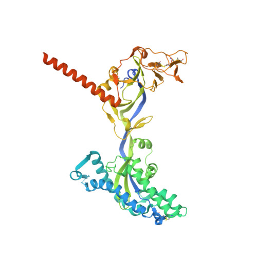
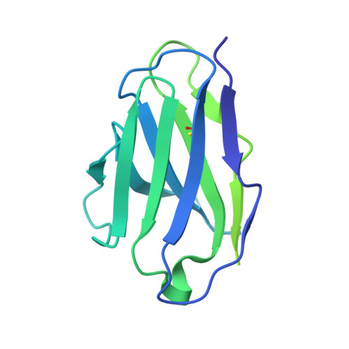
    Hendra virus (HeV) and Nipah virus (NiV) are henipaviruses (HNVs) causing respiratory illness and severe encephalitis in humans, with fatality rates of 50-100%. There are no licensed therapeutics or vaccines to protect humans. HeV and NiV use a receptor-binding glycoprotein (G) and a fusion glycoprotein (F) to enter host cells. HNV F and G are the main targets of the humoral immune response, and the presence of neutralizing antibodies is a correlate of protection against NiV and HeV in experimentally infected animals. We describe here two cross-reactive F-specific antibodies, 1F5 and 12B2, that neutralize NiV and HeV through inhibition of membrane fusion. Cryo-electron microscopy structures reveal that 1F5 and 12B2 recognize distinct prefusion-specific, conserved quaternary epitopes and lock F in its prefusion conformation. We provide proof-of-concept for using antibody cocktails for neutralizing NiV and HeV and define a roadmap for developing effective countermeasures against these highly pathogenic viruses.


  • Organizational Affiliation
    • Department of Biochemistry, University of Washington, Seattle, WA, USA.

Macromolecule Content 

  • Total Structure Weight: 404.42 kDa 
  • Atom Count: 15,738 
  • Modeled Residue Count: 2,010 
  • Deposited Residue Count: 3,600 
  • Unique protein chains: 3

Macromolecules

Find similar proteins by:|  3D Structure
Entity ID: 1
MoleculeChains  Sequence LengthOrganismDetailsImage
Fusion glycoprotein F0A,
D [auth B],
G [auth E]
543Henipavirus nipahenseMutation(s): 0 
UniProt
Find proteins for Q9IH63 (Nipah virus)
Explore Q9IH63 
Go to UniProtKB:  Q9IH63
Entity Groups
Sequence Clusters30% Identity50% Identity70% Identity90% Identity95% Identity100% Identity
UniProt GroupQ9IH63
Glycosylation
Glycosylation Sites: 4
Sequence Annotations
Expand
Reference Sequence
Find similar proteins by:|  3D Structure
Entity ID: 2
MoleculeChains  Sequence LengthOrganismDetailsImage
12B2 Fab light chainB [auth L],
E [auth C],
H [auth F]
214Mus musculusMutation(s): 0 
Find similar proteins by:|  3D Structure
Entity ID: 3
MoleculeChains  Sequence LengthOrganismDetailsImage
12B2 heavy chainC [auth H],
F [auth D],
I [auth G]
443Mus musculusMutation(s): 0 

Oligosaccharides

Help  
Entity ID: 4
MoleculeChains Length2D Diagram GlycosylationD Interactions
2-acetamido-2-deoxy-beta-D-glucopyranose-(1-4)-2-acetamido-2-deoxy-beta-D-glucopyranose
J [auth I],
L [auth K],
N,
P,
R,
J [auth I],
L [auth K],
N,
P,
R,
T
2N-Glycosylation
Glycosylation Resources
GlyTouCan: G42666HT
GlyCosmos: G42666HT
GlyGen: G42666HT
Entity ID: 5
MoleculeChains Length2D Diagram GlycosylationD Interactions
beta-D-mannopyranose-(1-4)-2-acetamido-2-deoxy-beta-D-glucopyranose-(1-4)-2-acetamido-2-deoxy-beta-D-glucopyranoseK [auth J],
O,
S
3N-Glycosylation
Glycosylation Resources
GlyTouCan: G15407YE
GlyCosmos: G15407YE
GlyGen: G15407YE
Entity ID: 6
MoleculeChains Length2D Diagram GlycosylationD Interactions
alpha-D-mannopyranose-(1-3)-[alpha-D-mannopyranose-(1-6)]beta-D-mannopyranose-(1-4)-2-acetamido-2-deoxy-beta-D-glucopyranose-(1-4)-[alpha-L-fucopyranose-(1-6)]2-acetamido-2-deoxy-beta-D-glucopyranose
M, Q, U
6N-Glycosylation
Glycosylation Resources
GlyTouCan: G82348BZ
GlyCosmos: G82348BZ
GlyGen: G82348BZ

Experimental Data & Validation

Experimental Data

  • Method: ELECTRON MICROSCOPY
  • Resolution: 2.90 Å
  • Aggregation State: PARTICLE 
  • Reconstruction Method: SINGLE PARTICLE 
EM Software:
TaskSoftware PackageVersion
RECONSTRUCTIONcryoSPARC

Structure Validation

View Full Validation Report


Entry History 

& Funding Information

Deposition Data


Funding OrganizationLocationGrant Number
National Institutes of Health/National Institute Of Allergy and Infectious Diseases (NIH/NIAID)United StatesDP1AI158186
National Institutes of Health/National Institute of General Medical Sciences (NIH/NIGMS)United StatesR01GM120553

Revision History  (Full details and data files)

  • Version 1.0: 2021-05-05
    Type: Initial release
  • Version 1.1: 2021-05-12
    Changes: Database references
  • Version 1.2: 2021-05-26
    Changes: Database references
  • Version 1.3: 2024-11-13
    Changes: Data collection, Database references, Structure summary