7KG3 | pdb_00007kg3

Crystal structure of CoV-2 Nsp3 Macrodomain


Experimental Data Snapshot

  • Method: X-RAY DIFFRACTION
  • Resolution: 1.45 Å
  • R-Value Free: 
    0.177 (Depositor), 0.179 (DCC) 
  • R-Value Work: 
    0.158 (Depositor), 0.158 (DCC) 
  • R-Value Observed: 
    0.158 (Depositor) 

Starting Model: experimental
View more details

wwPDB Validation 3D Report Full Report

Validation slider image for 7KG3

Ligand Structure Quality Assessment 


This is version 1.4 of the entry. See complete history

Literature

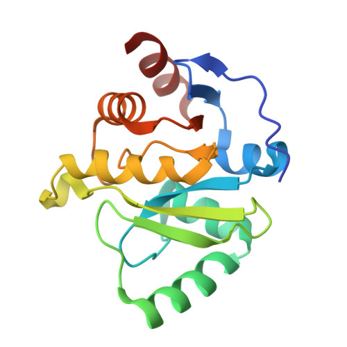
Targeting SARS-CoV-2 Nsp3 macrodomain structure with insights from human poly(ADP-ribose) glycohydrolase (PARG) structures with inhibitors.

Brosey, C.A.Houl, J.H.Katsonis, P.Balapiti-Modarage, L.P.F.Bommagani, S.Arvai, A.Moiani, D.Bacolla, A.Link, T.Warden, L.S.Lichtarge, O.Jones, D.E.Ahmed, Z.Tainer, J.A.

(2021) Prog Biophys Mol Biol 163: 171-186

  • DOI: https://doi.org/10.1016/j.pbiomolbio.2021.02.002
  • Primary Citation Related Structures: 
    7KFP, 7KG0, 7KG1, 7KG3, 7KG6, 7KG7, 7KG8, 7KXB, 7LG7

  • PubMed Abstract: 

    Arrival of the novel SARS-CoV-2 has launched a worldwide effort to identify both pre-approved and novel therapeutics targeting the viral proteome, highlighting the urgent need for efficient drug discovery strategies. Even with effective vaccines, infection is possible, and at-risk populations would benefit from effective drug compounds that reduce the lethality and lasting damage of COVID-19 infection. The CoV-2 MacroD-like macrodomain (Mac1) is implicated in viral pathogenicity by disrupting host innate immunity through its mono (ADP-ribosyl) hydrolase activity, making it a prime target for antiviral therapy. We therefore solved the structure of CoV-2 Mac1 from non-structural protein 3 (Nsp3) and applied structural and sequence-based genetic tracing, including newly determined A. pompejana MacroD2 and GDAP2 amino acid sequences, to compare and contrast CoV-2 Mac1 with the functionally related human DNA-damage signaling factor poly (ADP-ribose) glycohydrolase (PARG). Previously, identified targetable features of the PARG active site allowed us to develop a pharmacologically useful PARG inhibitor (PARGi). Here, we developed a focused chemical library and determined 6 novel PARGi X-ray crystal structures for comparative analysis. We applied this knowledge to discovery of CoV-2 Mac1 inhibitors by combining computation and structural analysis to identify PARGi fragments with potential to bind the distal-ribose and adenosyl pockets of the CoV-2 Mac1 active site. Scaffold development of these PARGi fragments has yielded two novel compounds, PARG-345 and PARG-329, that crystallize within the Mac1 active site, providing critical structure-activity data and a pathway for inhibitor optimization. The reported structural findings demonstrate ways to harness our PARGi synthesis and characterization pipeline to develop CoV-2 Mac1 inhibitors targeting the ADP-ribose active site. Together, these structural and computational analyses reveal a path for accelerating development of antiviral therapeutics from pre-existing drug optimization pipelines.


  • Organizational Affiliation
    • Department of Molecular and Cellular Oncology, M. D. Anderson Cancer Center, Houston, TX, 77030, USA. Electronic address: CABrosey@mdanderson.org.

Macromolecule Content 

  • Total Structure Weight: 18.89 kDa 
  • Atom Count: 1,493 
  • Modeled Residue Count: 166 
  • Deposited Residue Count: 169 
  • Unique protein chains: 1

Macromolecules

Find similar proteins by:|  3D Structure
Entity ID: 1
MoleculeChains  Sequence LengthOrganismDetailsImage
Non-structural protein 3169Severe acute respiratory syndrome coronavirus 2Mutation(s): 0 
Gene Names: rep1a-1b
EC: 3.4.22
UniProt
Find proteins for P0DTD1 (Severe acute respiratory syndrome coronavirus 2)
Explore P0DTD1 
Go to UniProtKB:  P0DTD1
Entity Groups
Sequence Clusters30% Identity50% Identity70% Identity90% Identity95% Identity100% Identity
UniProt GroupP0DTD1
Sequence Annotations
Expand
Reference Sequence

Small Molecules

Ligands 4 Unique
IDChains Name / Formula / InChI Key2D Diagram3D Interactions
MES
(Subject of Investigation/LOI)

Query on MES


Download:Ideal Coordinates CCD File
D [auth A]2-(N-MORPHOLINO)-ETHANESULFONIC ACID
C6 H13 N O4 S
SXGZJKUKBWWHRA-UHFFFAOYSA-N
MLI

Query on MLI


Download:Ideal Coordinates CCD File
B [auth A],
C [auth A]
MALONATE ION
C3 H2 O4
OFOBLEOULBTSOW-UHFFFAOYSA-L
SO4

Query on SO4


Download:Ideal Coordinates CCD File
F [auth A],
G [auth A],
H [auth A]
SULFATE ION
O4 S
QAOWNCQODCNURD-UHFFFAOYSA-L
GOL

Query on GOL


Download:Ideal Coordinates CCD File
E [auth A]GLYCEROL
C3 H8 O3
PEDCQBHIVMGVHV-UHFFFAOYSA-N

Experimental Data & Validation

Experimental Data

  • Method: X-RAY DIFFRACTION
  • Resolution: 1.45 Å
  • R-Value Free:  0.177 (Depositor), 0.179 (DCC) 
  • R-Value Work:  0.158 (Depositor), 0.158 (DCC) 
  • R-Value Observed: 0.158 (Depositor) 
Space Group: P 31 2 1
Unit Cell:
Length ( Å )Angle ( ˚ )
a = 112.594α = 90
b = 112.594β = 90
c = 41.407γ = 120
Software Package:
Software NamePurpose
PHENIXrefinement
XDSdata reduction
Aimlessdata scaling
PHASERphasing

Structure Validation

View Full Validation Report


Ligand Structure Quality Assessment 


Entry History 

& Funding Information

Deposition Data


Funding OrganizationLocationGrant Number
National Institutes of Health/National Cancer Institute (NIH/NCI)United StatesR01 CA200231

Revision History  (Full details and data files)

  • Version 1.0: 2020-10-28
    Type: Initial release
  • Version 1.1: 2021-01-27
    Changes: Structure summary
  • Version 1.2: 2021-03-10
    Changes: Database references
  • Version 1.3: 2021-06-16
    Changes: Database references
  • Version 1.4: 2023-10-18
    Changes: Data collection, Database references, Refinement description